43 brain stem labels
Human Brain Model for Neuroscience Teaching with Labels 2 Times Life ... CLEAR DISPLAY - BEAMNOVA two-times life-size Human Brain Model consists of 4 components, with laser engraved labels, perfect for students and clinicians. USER- FRIENDLY - All 4 pieces come apart for viewing, and easily fit back together like a puzzle. ... with a list of detailed anatomical structure annotation, allowing you to study deep within ... Brainstem - Wikipedia The brainstem (or brain stem) is the posterior stalk-like part of the brain that connects the cerebrum with the spinal cord. In the human brain the brainstem is composed of the midbrain, the pons, and the medulla oblongata.The midbrain is continuous with the thalamus of the diencephalon through the tentorial notch,: 152 and sometimes the diencephalon is included in the brainstem.
Brain Label (Remote) - The Biology Corner This brain labeling activity was created for remote learners as an alternative to the labeling and coloring worksheet we would traditionally do in class. Instead of coloring and labeling on printouts, students use google slides to drag labels to the images or type the answers into text boxes.

Brain stem labels
Brain Stem : Anatomy, Location & Function - Anatomy Info Brain Stem: The brainstem also has known as brain stem is the back part of the brain, joining and structurally continuous with the spinal cord. In the brain, brainstem comprises the midbrain, the pons, and the medulla oblongata. The brain stem performs the motor and sensory innervation to the face and neck through the cranial nerves. Brainstem Anatomy - W-Radiology The brainstem refers to the middle part of the brain (1). It consists of the medulla, pons, and midbrain. The brainstem helps relay sensory information, such as pain, eye and mouth movement, involuntary muscle movements, consciousness, respirations, hunger, and cardiac function (2). Brainstem: Overview, Function & Anatomy - Cleveland Clinic Brainstem Your brainstem is the bottom, stalklike portion of your brain. It connects your brain to your spinal cord. Your brainstem sends messages to the rest of your body to regulate balance, breathing, heart rate and more. Sudden injuries, and brain or heart conditions may affect how your brainstem works. Appointments 866.588.2264
Brain stem labels. Parts of the brain stem label diagram Quiz - purposegames.com Vertebra label quiz 7p Image Quiz. Respiratory System label quiz 16p Image Quiz. Bones, sutures and frontanelle of fetal skull 12p Image Quiz. Parts of the brain stem 16p Image Quiz. Structures & circumference of newborn skull 9p Image Quiz. Innominate Bone label quiz - the pelvis 14p Image Quiz. Male reproductive system label diagram 15p Image ... Brain Stem Parts Anatomical Model in Educational Labeled Outline ... Brain stem parts anatomical model in educational labeled outline diagram. Biological sections location with titles scheme vector illustration. Thalamus, midbrain, pons, medulla and spinal cord graph. brain stem, cord graph, vector illustration, scheme vector, vector, brain, educational, diagram, location, outline, stem, model, anatomical, thalamus, Brain: Atlas of human anatomy with MRI - e-Anatomy - IMAIOS The brain stem subdivided into midbrain, pons and medulla oblongata (bulb, myelencephalon). Brainstem. The cranial nerves at the level of their emergence, and the cranial nerve nuclei with a projection of their supposed position on the brain stem. The brain ventricles (lateral ventricles, third ventricle, fourth ventricle) with the choroid plexus. Retrograde labeling of neurones in the brain stem following ... by BE Jones · 1987 · Cited by 80 — After injections into the anterior medial frontal cortex, a small number of retrogradely labeled cells were found in the brain stem within the laterodorsal ...
The Brainstem - TeachMeAnatomy The midbrain (also known as the mesencephalon) is the most superior of the three regions of the brainstem. It acts as a conduit between the forebrain above and the pons and cerebellum below. The midbrain is the smallest of the three regions of the brainstem, measuring around 2cm in length. As it ascends, the midbrain travels through the opening ... Cross-sectional anatomy of the brain - e-Anatomy - IMAIOS Anatomy of the brain: how to view anatomical labels. This module is a comprehensive and affordable learning tool for medical students and residents and especially for neuroradiologists and radiation oncologists. It provides access to an atlas and to images in axial planes, allowing the user to learn and review neuroanatomy interactively. ... Brain Parts - Labels | Psychology Quiz - Quizizz Brain Parts - Labels DRAFT. 10th - 12th grade. 0 times. Social Studies. 0% average accuracy. 6 minutes ago. nfulle1. 0. Save. Edit. Edit. Brain Parts - Labels DRAFT. 6 minutes ago. by nfulle1. ... brain stem. Tags: Question 13 . SURVEY . 30 seconds . Q. This structure helps pass information between the two hemispheres of the brain. answer choices Anatomy of the Brain - Mayfield Clinic The brain is composed of the cerebrum, cerebellum, and brainstem (Fig. 1). A side view illustration of the human brain, with areas labeled and colored.
Diagram Of Brain with their Labelings and Detailed Explanation The parietal lobe is found at the upper back of our brain. This lobe functions by controlling all our complex behaviours, including senses of vision, the sense of touch, spatial orientation and body awareness. It manages body position, movements, the perception of stimuli, orientation, handwriting and visuospatial processing. The Occipital Lobe Amazon.com: Labeled Brain Model 1-16 of 586 results for "Labeled Brain Model" RESULTS Amazon's Choice Learning Resources Cross-section Brain Model - 2 Pieces, Ages 7+ Brain Anatomy Model, Brain Functions Model, Human Anatomy for Kids, Foam Brain Model 1,391 $18 88 $19.99 Get it as soon as Mon, Mar 28 FREE Shipping on orders over $25 shipped by Amazon More Buying Choices Labeled Diagrams of the Human Brain You'll Want to Copy Now Labeled Diagrams of the Human Brain Central Core The central core consists of the thalamus, pons, cerebellum, reticular formation and medulla. These five regions are the central areas that regulate breathing, pulse, arousal, balance, sleep and early stages of processing sensory information. University of Wisconsin-Madison University of Wisconsin-Madison
Anatomical Foundations of Neuroscience
3D Brain This interactive brain model is powered by the Wellcome Trust and developed by Matt Wimsatt and Jack Simpson; reviewed by John Morrison, Patrick Hof, and Edward Lein. Structure descriptions were written by Levi Gadye and Alexis Wnuk and Jane Roskams .

Nervous System
Brain Anatomy - Brainlab.org The midbrain consists of various cranial nerve nuclei, tectum, tegmentum, colliculi, and crura cerebi. The hindbrain, also referred to as the brainstem, is made ...
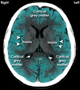
Brain CT - NeurologyNeeds.com
Brain Models - San Diego Mesa College NERVOUS SYSTEM - BRAIN MODELS ... Anterior Brainstem · Posterior Brainstem · Medial Brainstem · Half-Head · Sagittal Section ... Brain Models Labeled.
11 Best Images of Nervous System Labeling Worksheets - NAME LAB TIME ...
brainstem | Definition, Structure, & Function | Britannica brainstem, area at the base of the brain that lies between the deep structures of the cerebral hemispheres and the cervical spinal cord and that serves a critical role in regulating certain involuntary actions of the body, including heartbeat and breathing. The brainstem is divided into three sections in humans: the midbrain (mesencephalon), the pons (metencephalon), and the medulla oblongata ...

Tres aplicaciones gratis de iPad para apoyar tus clases de ciencia
Neuroanatomy: Brainstem Cross Sections - Anatomy Guy 2012-06-08 Brainstem Sections Handout

WMU Psychology Department: Lisa Baker
MS Brain Lesions and Their Effects - Verywell Health Your healthcare provider may identify a brainstem lesion based on your history and physical examination. 3 However, because many of the signs and symptoms of brainstem lesions in MS are the same as those of other MS lesions, diagnostic tests can help identify where your MS lesions are located.

The brain - structure and function - Cancer Information - Macmillan ...
Brainstem: Anatomy, Function, and Treatment - Verywell Health The brainstem is a stem shaped structure, extending down from the posterior (back) part of the brain to the spinal cord. It is protected by the meninges, which are composed of three layers of sheet-like connective tissue that envelop the brain and spinal cord. Outside the meninges, the brainstem is shielded by the lower part of the skull.
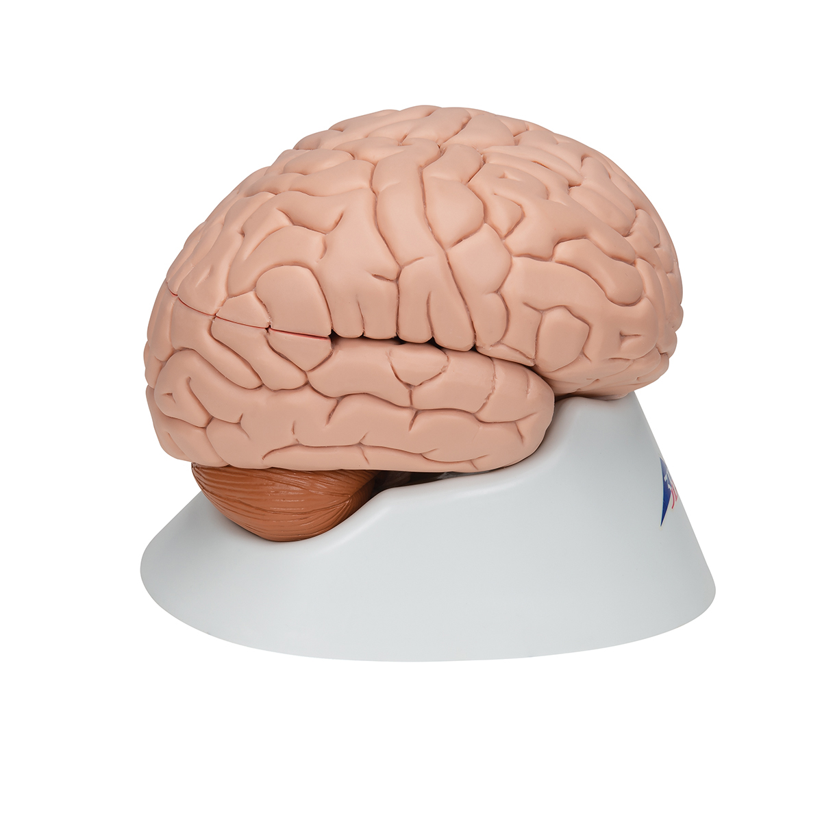
Anatomical Teaching Models | Plastic Human Brain Models | 8-Part Brain ...
brain model labeled - Amazon.com Results 1 - 16 of 432 — Ultrassist Brain Model for Neuroscience, Brain Model Labeled with ... of Brain Stem | Half of Cerebellum | Instruction Manual Included.
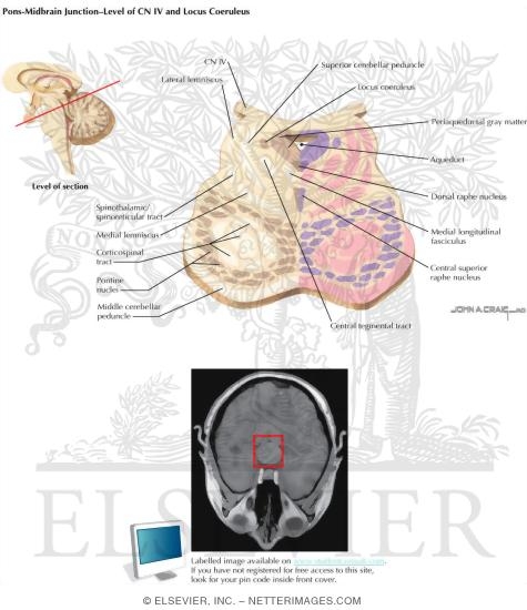
Pons-Midbrain Junction - Level of CN IV and Locus Coeruleus
Labeling the Brain | Human Anatomy Quiz - Quizizz Play this game to review Human Anatomy. What lobe is orange in this picture?
Post a Comment for "43 brain stem labels"