44 labelled diagram of compound microscope
Draw a neat labelled diagram of a compound microscope class 12 physics CBSE Compound Microscope Book Online Demo Answer Draw a neat labelled diagram of a compound microscope. Derive the magnifying power for it. A telescope has an objective of focal length 140cm and an eyepiece of focal length 5cm. Find the magnifying power and separation between objective and eyepiece. Answer Verified 156.3k + views 1 likes (i) Draw a neat labelled diagram of a compound microscope explain ... Thus the final image A'' B'' formed by the microscope is inverted and magnified and its position is outside the objective and eyepiece towards objective lens. Magnifying power of compound microscope is for final image at distance of distinct vision for final image at infinity (ii) For large magnifying power, f0 and fe both have to be small.
Draw a Labelled Ray Diagram Showing the Formation of Image by a ... Diagram Draw a labelled ray diagram showing the formation of image by a compound microscope in normal adjustment. Derive the expression for its magnifying power. Advertisement Remove all ads Solution A simple microscope has a limited maximum magnification (≤ 9) for realistic focal lengths.

Labelled diagram of compound microscope
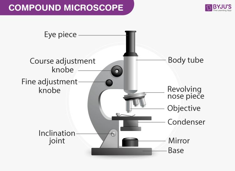
Labelled Diagram of Compound Microscope - Biology Discussion The below mentioned article provides a labelled diagram of compound microscope. Part # 1. The Stand: The stand is made up of a heavy foot which carries a curved inclinable limb or arm bearing the body tube. The foot is generally horse shoe-shaped structure (Fig. 2) which rests on table top or any other surface on which the microscope in kept. Parts of Stereo Microscope (Dissecting microscope) - labeled diagram ... If you would like to learn optical components of a compound microscope, please visit Compound Microscope Parts - Labeled Diagram and their Functions, and this article. How to use a stereo (dissecting) microscope. Follow these steps to put your stereo microscopes in work: 1. Set your microscope on a tabletop or other flat sturdy surface where ... Compound microscope - their parts and function - Microscopy4kids Labeled diagram of a compound microscope. Optical components of a compound microscope. The term "compound" refers to the microscope having more than one lens. Compound microscopes generate magnified images through an aligned pair of the objective lens and the ocular lens. In contrast, "simple microscopes" have only one convex lens and ...
Labelled diagram of compound microscope. Draw a neat labelled diagram of a compound microscope and explain its ... Dividing and multiplying by I1 G1 on the right side, we get Magnifying power of the objective (m0) = I1G1/OJ = Height of the image due to the objective. Magnifying power of the eye piece (me) = IG/I1G1 = Height of the final image / Height of the object for the eyepiece. ∴ m = m0 × me ..... (1) Microscope Parts and Functions With Labeled Diagram and Functions How ... Coarse adjustment: Brings the specimen into general focus. Fine adjustment: Fine tunes the focus and increases the detail of the specimen. Nosepiece: A rotating turret that houses the objective lenses. The viewer spins the nosepiece to select different objective lenses. Objective lenses: One of the most important parts of a compound microscope ... Compound Microscope- Definition, Labeled Diagram, Principle, Parts, Uses A compound light microscope mostly uses a low voltage bulb as an illuminator. The stage is the flat platform where the slide is placed. Nosepiece and Aperture Nosepiece is a rotating turret that holds the objective lenses. The viewer spins the nosepiece to select different objective lenses. Parts of a microscope with functions and labeled diagram Q. List down the 18 parts of a Microscope. 1. Ocular Lens (Eye Piece) 2. Diopter Adjustment 3. Head 4. Nose Piece 5. Objective Lens 6. Arm (Carrying Handle) 7. Mechanical Stage 8. Stage Clip 9. Aperture 10. Diaphragm 11. Condenser 12. Coarse Adjustment 13. Fine Adjustment 14. Illuminator (Light Source) 15. Stage Controls 16. Base 17.
Microscope Parts, Function, & Labeled Diagram - slidingmotion Condenser. The condenser is to focus the light, which passes from the microscopic illuminator to the specimen. This condenser is located just below the diaphragm. This diaphragm is one of the important parts of the compound microscope which will help to get an accurate and sharp image. The condenser has a magnification power of 400X and above. Compound Microscope Parts, Functions, and Labeled Diagram Nov 18, 2020 · Compound Microscope Parts, Functions, and Labeled Diagram Parts of a Compound Microscope Each part of the compound microscope serves its own unique function, with each being important to the function of the scope as a whole. Draw a ray diagram of compound microscope, when final image is formed ... Draw a ray diagram of compound microscope, when final image is formed at the minimum distance of distinct vision. Easy Solution Verified by Toppr It consist of two convex lenses, one objective of very small focal length with short aperture. And one Eyepiece with moderate focal length and large aperture. Compound Microscope - Diagram (Parts labelled), Principle and Uses Feb 03, 2022 · See: Labeled Diagram showing differences between compound and simple microscope parts Structural Components The three structural components include 1. Head This is the upper part of the microscope that houses the optical parts 2. Arm This part connects the head with the base and provides stability to the microscope.
Compound Microscope Parts - Labeled Diagram and their Functions - Rs ... Labeled diagram of a compound microscope Major structural parts of a compound microscope There are three major structural parts of a compound microscope. The head includes the upper part of the microscope, which houses the most critical optical components, and the eyepiece tube of the microscope. Parts of a Compound Microscope and Their Functions Compound microscope magnification is determined by multiplying the eyepiece and objective powers. When viewed through a 5X eyepiece with a 10X objective, an item is magnified 5 x 10=50 times. The magnification is 10 x 45 = 450 times when using a 10X eyepiece and a 45X objective. How to Use the Compound Microscope Draw a labelled ray diagram of an image formed by a class 12 physics CBSE Complete step by step answer: The ray diagram an image formed by a compound microscope, when the final image lies at the least distance of distinct vision (D) is as follows: In the above ray diagram, O is the objective lens and E is the eyepiece lens.In the above ray diagram of the compound microscope, AB is the object, A'B' is the image ... Solved VIN Draw the labelled diagram of compound microscope | Chegg.com VIN Draw the labelled diagram of compound microscope when image is formed at infinity. A microscope consisting of two convex lenses of focal lengths 2 cm and 5 cm are placed 20 cm apart. Where must the object be placed so that the final image is formed at infinity?
Labelled Diagram Of A Tick - Top Label Maker
Draw a labelled ray diagram of a compound microscope. Explain its ... (a) Draw a labelled ray diagram showing the formation of a final image by a compound microscope at least distance of distinct vision. (b) The total magnification produced by a compound microscope is 20. The magnification produced by the piece is 5. The microscope is focussed on a certain object.

Clipart Panda - Free Clipart Images
BYJUS BYJUS

Image result for compound light microscope parts and their functions ...
Microscope Types (with labeled diagrams) and Functions Compound microscope labeled diagram Compound microscope functions: It finds great application in areas of pathology, pedology, forensics etc Its greater order of magnification allows for deeper study of microbial organisms to Detect the cause of diseases Study the mineral composition in soils

Microscope Labeling
(a) Draw a labelled ray diagram of a compound microscope. (b) Derive an ... selected May 26, 2018 by Vikash Kumar Best answer (a) Labelled diagram of compound microscope. The objective lens form image A' B' near the first focal point ofeyepiece. (b) Angular magnification of objective lens m0 = linear magnification h'/h where L is the distance between second focal point of the objective and first focal point of eyepiece.
Microscope Review.wmv - YouTube
(a) Draw the labelled ray diagram for the formation of image by a ... (a) Draw the labelled ray diagram for the formation of image by a compound microscope. Derive an expression for its total magnification (or magnifying power), when the final image is formed at the near point. (b) Why both objective and eyepiece of a compound microscope must have short focal lengths?

Cell Theory timeline | Timetoast timelines
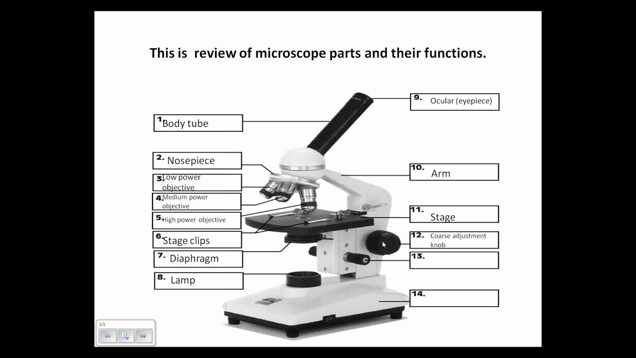
COMPOUND MICROSCOPE - Labelled diagram eyepiece , coarse adjustment knob, fine adjustment knob, objectives, stage, arm, mirror, base, draw tube, condenser, body tube, iris diaphragm .
MP Board Class 10 Science 2018 Previous Year Question Paper Solutions ...
Draw a ray diagram of a compound microscope. Write the expression for ... Ray diagram of a compound microscope.When the final image is formed at the least distance of distinct vision,For the image formed at infinity, ue = feand By making focal length of the objective small, the magnifying power can be increased. Ray diagram of a compound microscope.When the final image is formed at the least distance of distinct ...

The History of Microscopes timeline | Timetoast timelines
Compound Microscope Labeled Diagram | Quizlet QUESTION. The total magnification of a specimen being viewed with a 10X ocular lens and a 40X objective lens is. 15 answers. QUESTION. a mosquito beats its wings up and down 600 times per second, which you hear as a very annoying 600 Hz sound. if the air outside is 20 C, how far would a sound wave travel between wing beats. 2 answers.

Microscope Labeling Game
draw the labelled ray diagram for the formation of image by a compound ... A compound microscope consists of an objective of focal length 1 cm and eye piece of focal length 5 c separated by 12.2 cm. At what distance from the objective should an object be placed so that the final image is formed at least distance of distinct vision? A convex lens of focal length 15 cm is placed in front of a convex mirror.
Post a Comment for "44 labelled diagram of compound microscope"