43 label the structure of the muscle fiber
Label structure of skeletal muscle Diagram | Quizlet a long, filamentous organelle found within muscle cells that has a banded appearance tendon cordlike extension of connective tissue beyond the muscle, serving to attach it to the bone sarcolemma plasma membrane of a muscle cell epimysium just deep to the deep fascia (surrounds entire muscle) perimysium Muscle Fiber Anatomy Quiz - PurposeGames.com This is an online quiz called Muscle Fiber Anatomy. There is a printable worksheet available for download here so you can take the quiz with pen and paper. Your Skills & Rank. Total Points. 0. Get started! Today's Rank--0. Today 's Points. One of us! Game Points. 15. You need to get 100% to score the 15 points available.
General Anatomy of Skeletal Muscle Fibers - GetBodySmart Skeletal Muscle Fiber Location and Arrangement. are located inside muscles, where they are organized into bundles called […] Internal Anatomy of Skeletal Muscle Fibers. An interactive quiz about the internal anatomy of skeletal muscle fibers, featuring illustrations-based multiple choice questions.
Label the structure of the muscle fiber
› image › nervovNervous System: Explore the Nerves with Interactive Anatomy ... Nov 02, 2020 · Efferent neurons (also called motor neurons) carry signals from the gray matter of the CNS through the nerves of the peripheral nervous system to effector cells. The effector may be smooth, cardiac, or skeletal muscle tissue or glandular tissue. The effector then releases a hormone or moves a part of the body to respond to the stimulus. Ultrastructure of Muscle - Skeletal - Sliding Filament - TeachMeAnatomy Ultrastructural Appearance of Skeletal Muscle. The striated appearance of skeletal muscle fibres is due to the organisation of two contractile proteins: actin (thin filament) and myosin (thick filament). The functional unit of contraction in a skeletal muscle fibre is the sarcomere, which runs from Z line to Z line. Solved A. Labeling Label the structure of the muscle fiber | Chegg.com Answer A. Label Myofibrils Sarcoplasmic reticulum Terminal cisrernae T Tubule (Transverse t … View the full answer Transcribed image text: A. Labeling Label the structure of the muscle fiber Sarca Plasmic retiulom tencian Cistor T-woie triad Sarcomere 3. 10 4. 11 5 6 7. Sarco leme 8 9 Sin Ptis Vesiick 10. 11. 12. Aton B. Matching on the right.
Label the structure of the muscle fiber. › tag › anatomyAnatomy quizzes - PurposeGames Brain Structure Identification by KeeneyQuest 151,253 plays ... Label the Muscles by annamalcracker 50,409 plays ... Skeletal muscle fiber model by cwalsh2 35,847 ... anatomyzone.comAnatomyZone - Your Guide to Human Anatomy AnatomyZone is the leading resource for simple and concise 3D anatomy tutorials, with over 200 videos and a new range of interactive 3D anatomy models. en.wikipedia.org › wiki › Dietary_fiberDietary fiber - Wikipedia Dietary fiber (British spelling fibre) or roughage is the portion of plant-derived food that cannot be completely broken down by human digestive enzymes. Dietary fibers are diverse in chemical composition, and can be grouped generally by their solubility, viscosity, and fermentability, which affect how fibers are processed in the body. Art-labeling Activity: The Structure of a Skeletal Muscle Fiber Start studying Art-labeling Activity: The Structure of a Skeletal Muscle Fiber. Learn vocabulary, terms, and more with flashcards, games, and other study tools. ... Test. PLAY. Match. Created by. BabeRuthless0504. Terms in this set (2) Art-labeling Activity: The Structure of a Skeletal Muscle Fiber... Art-labeling Activity: The Structure of a ...
Skeletal Muscle Fiber Labeling Flashcards | Quizlet dark a band light i band nucleus purple dots on muscle fiber thin (actin) filament thick (myosin) filament z disc h zone m line elastic (titin) filament muscle fiber a single muscle cell can also be called ... myofibril there are many __________ in a single muscle fiber/cell. sarcomere there are many ____________ in a single myofibril. Muscle Fiber Labeling Quiz - PurposeGames.com This is an online quiz called Muscle Fiber Labeling Quiz. There is a printable worksheet available for download here so you can take the quiz with pen and paper. Your Skills & Rank. Total Points. 0. Get started! ... Bone Structure (Compact Bone) 10p Image Quiz. PurposeGames Create. Play. Learn. DOCX Label the diagram of the skeletal muscle with the following terms ... Label the diagram of the muscle fiber below with the following terms: actin (thin filaments), neuromuscular junction, myofibril, myosin (thick filaments), sarcolemma, sarcomere, sarcoplasmic reticulum, T-tubule, Z-line. 3. Color the muscles of the head and neck in the diagrams below. 4. Color the muscles of the upper back and chest in the ... › nutrition › healthiest-beansThe 9 Healthiest Beans and Legumes You Can Eat May 04, 2022 · Also known as garbanzo beans, chickpeas are a great source of fiber and protein. One cup (164 grams) of cooked chickpeas contains ():Calories: 269 Protein: 14.5 grams Fat: 4.25 grams Carbohydrates ...
Correctly Label The Following Parts Of A Skeletal Muscle Fiber A skeletal muscle fiber is composed of a plasma membrane and a specialized smooth endoplasmic reticulum. It also contains sarcomeres and calcium ions. In addition to the plasma membrane, a skeletal muscle fiber has numerous myofibrils. During a contraction, the force is transmitted through the tendon to the bone, producing a skeletal movement. › blog › health-benefits-ofHealth Benefits of Bread | Is Bread Good For You? Feb 17, 2021 · 1. Bread Contains Fiber. Different bread products contain varying amounts of fiber, but you can generally find at least 1 gram per serving. And this is an essential nutrient to eat every day. Many know fiber for keeping your digestive system moving, but it does more than that. When you eat fiber, it can help you: What is the structure of a muscle fiber? - AskingLot.com What is the structure of a muscle fiber? Muscle Fiber. A skeletal muscle fiber is surrounded by a plasma membrane called the sarcolemma, which contains sarcoplasm, the cytoplasm of muscle cells. A muscle fiber is composed of many fibrils, which give the cell its striated appearance. Click to see full answer. Label structure of skeletal muscle Diagram | Quizlet Start studying Label structure of skeletal muscle. Learn vocabulary, terms, and more with flashcards, games, and other study tools.
Microscopic structure of skeletal muscle by Dr. S. N. Singh
Answered: Label the structure of the muscle… | bartleby Solution for Label the structure of the muscle fiber. close. Start your trial now! First week only $4.99! arrow_forward. learn. write. tutor. study resourcesexpand_more. Study Resources. We've got the study and writing resources you need for your assignments. Start exploring! Subjectschevron_right; RESOURCES ...

How Heat Affects Muscle Fibers in Meat | ThermoWorks
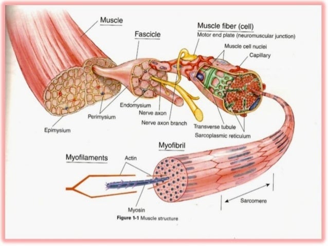
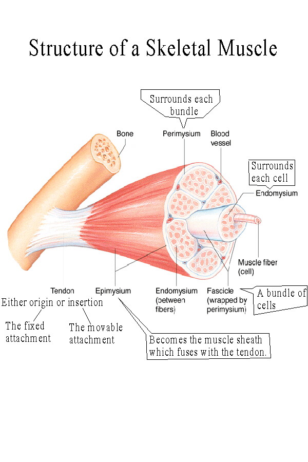
10.2 Skeletal Muscle - Anatomy & Physiology Figure 10.2.1 - The Three Connective Tissue Layers: Bundles of muscle fibers, called fascicles, are covered by the perimysium. Muscle fibers are covered by the endomysium. Inside each skeletal muscle, muscle fibers are organized into bundles, called fascicles, surrounded by a middle layer of connective tissue called the perimysium.
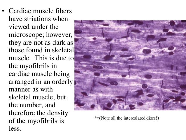
3. cardiac muscle tissue
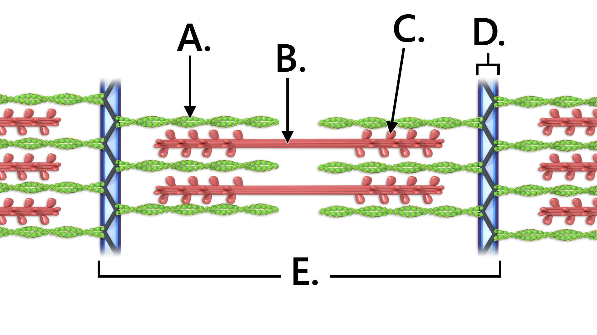
Structures of the Skeletal Muscle Fiber Flashcards | Quizlet -Muscle fibers are filled with threads called myofibrils separated by SR (sarcoplasmic reticulum) -Myofilaments (thick & thin filaments) are the contractile proteins of muscle that actually cause muscles to contract. Sarcoplasmic Reticulum (SR) -Stores Ca+2 in a relaxed muscle -Release of Ca+2 triggers muscle contraction Filaments and the Sarcomere

A & P Ch 6 Musclular System Student PPT
Art-labeling activity: structure of skeletal muscle fiber. Drag the ... Art-Labeling Activity: The Structure Of A Sarcomere Part A Drag The Labels To The Appropriate Location In The Figure. Reset Help A Band Barmere Hand Band MI Art-Labeling Activity: The Structure Of A Skeletal Muscle Fiber Part A Drag The Labels Onto...

SMOOTH MUSCLE | Microanatomy Web Atlas | Gwen V. Childs, Ph.D.
› pmc › articlesSarcopenia: Aging-Related Loss of Muscle Mass and Function Jan 01, 2019 · A. Methodological Problems in the Study of Aging Skeletal Muscle. The study of sarcopenia in humans is complicated by the long duration of the aging process, large variability among individuals, and multiple factors affecting muscle that are not primarily related to aging per se. Studies of aging can be conducted using either a cross-sectional or longitudinal design, but neither are free from ...
All About Muscle Growth | Precision Nutrition
UCSD Muscle Physiology Homepage - Fiber Structure A myofiber is a multinucleated single muscle cell (see Figure 1, below). Physically, they range in size from a under a hundred microns in diameter and a few millimeters in length to a few hundred microns across and a few centimeters in length. The cell is densely packed with contractile proteins, energy stores and signaling mechanisms.

Pin on Education: Anatomy
Muscle Fibers: Anatomy, Function, and More - Healthline Muscle tissue contains something called muscle fibers. Muscle fibers consist of a single muscle cell. They help to control the physical forces within the body. When grouped together, they can...
Skin Label Stock Illustration - Image: 46887482
Answered: Label the structure of the muscle fiber | bartleby Label the structure of the muscle fiber Transcribed Image Text: Review & Practice Sheet Section A Part A Label the structure of the muscle fiber. Reset Help Muscle fiber I tubule Sarcoplasmic Sarcolemma reticulum Axon Terminal cisterna Synaptic cleft Sarcomere Triad Synaptic vesicle Junctional Fold Perimysium Expert Solution
Skeletal Muscle Physiology Review Sheet | Clare Hays Biology Homepage
Solved A. Labeling Label the structure of the muscle fiber - Chegg Question: A. Labeling Label the structure of the muscle fiber 2. 10 3. 5. 6. 12 8. 10. 12 This problem has been solved! See the answer Show transcribed image text Expert Answer 100% (8 ratings) 1. Myofibril 2. Sarcoplasmic reticulum 3. Terminal cisternae 4.T- tubule 5. Triad ( a T-tubule surrounded by two terminal cisternae is called traid ) 6.
Post a Comment for "43 label the structure of the muscle fiber"