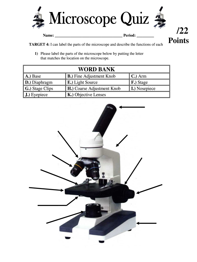
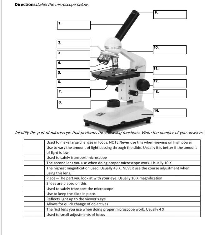
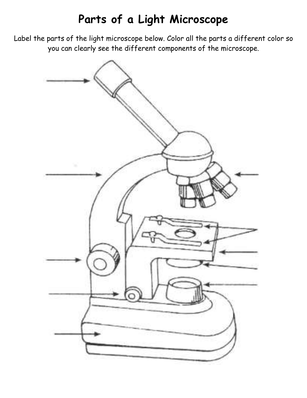
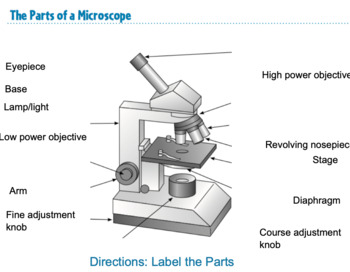
40 label the different parts of the microscope
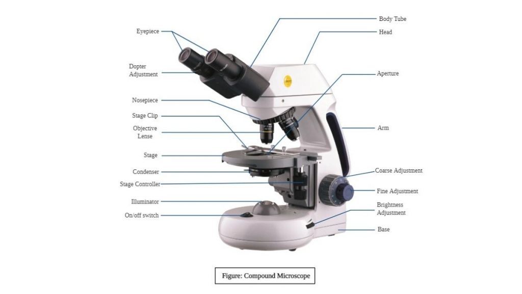
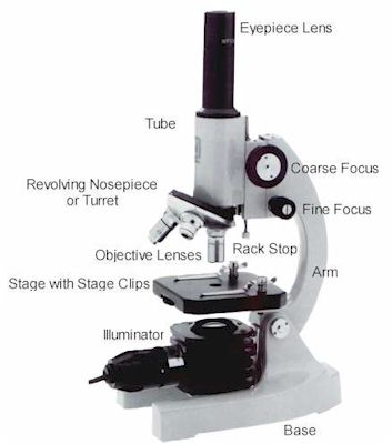
Parts of a microscope with functions and labeled diagram 19.4.2022 · Thank you very much it really helped me with my science home work since i in 8th grade and this my home work to draw a microscope label all the parts and the function thank may the holy father of holy ... This note is really helpeful to me to differet ways to different methology. Reply. Ansariasif. January 1, 2022 at 4:38 PM . Thanks ... Microscope, Microscope Parts, Labeled Diagram, and Functions 19.1.2022 · Revolving Nosepiece or Turret: Turret is the part of the microscope that holds two or multiple objective lenses and helps to rotate objective lenses and also helps to easily change power. Objective Lenses: Three are 3 or 4 objective lenses on a microscope. The objective lenses almost always consist of 4x, 10x, 40x and 100x powers. The most common eyepiece …
Guide to Teaching Kids About Cells | Science Explorers 25.4.2019 · To understand the parts of cells and what they do, first, teach kids about cells and what they are. Don’t be afraid to incorporate the scientific names for the parts of cells into your discussion. Repeating these names and the cell types will help to learn them. Young kids can learn the names of cell parts, too, even if they are complex.
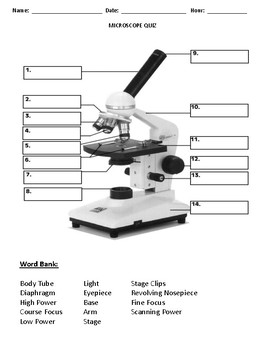
Label the different parts of the microscope
What is a Compound Microscope? | Microscope World Blog The Parts & Function of a Compound Microscope A compound microscope is a high power (high magnification) microscope that uses a compound lens system. A compound microscope has multiple lenses: the objective lens (typically 4x, 10x, 40x or 100x) is compounded (multiplied) by the eyepiece lens (typically 10x) to obtain a high magnification of 40x, 100x, 400x and 1000x. sciencing.com › difference-between-compoundDifference Between Compound & Dissecting Microscopes Mar 10, 2018 · The dissecting microscope is also known as a stereomicroscope. Because it has a long working distance, between 25 and 150 mm, it has a lower magnification ability. This gives the user the option to manipulate the specimen, even performing small dissections under the microscope. Live specimens can also be observed. › p-3470-what-is-aWhat is a Compound Microscope? | Microscope World Blog The Parts & Function of a Compound Microscope A compound microscope is a high power (high magnification) microscope that uses a compound lens system. A compound microscope has multiple lenses: the objective lens (typically 4x, 10x, 40x or 100x) is compounded (multiplied) by the eyepiece lens (typically 10x) to obtain a high magnification of 40x ...
Label the different parts of the microscope. depts.washington.edu › vurchinWelcome to Virtual Urchin - University of Washington 1. microscope basics. microscope tutorial: microscope measurement: ... (and only in Parts 1-3 of that activity). We're working to expand the choices. If you choose a ... microbenotes.com › parts-of-a-microscopeParts of a microscope with functions and labeled diagram Apr 19, 2022 · Figure: Diagram of parts of a microscope. There are three structural parts of the microscope i.e. head, base, and arm. Head – This is also known as the body. It carries the optical parts in the upper part of the microscope. Base – It acts as microscopes support. It also carries microscopic illuminators. Explanation and Labelled Images - New York Microscope Company 16.12.2020 · The Characteristics of a Fluorescence Microscope. The main parts of a fluorescent microscope overlap with the traditional light microscope. However, there are two main features that sets fluorescent microscope apart from the traditional microscope. One is the type of light source and the other is the use of specialized filter elements. › 6-label-the-microscopeLabel the microscope — Science Learning Hub Jun 08, 2018 · All microscopes share features in common. In this interactive, you can label the different parts of a microscope. Use this with the Microscope parts activity to help students identify and label the main parts of a microscope and then describe their functions. Drag and drop the text labels onto the microscope diagram. If you want to redo an ...
Microscope slide - Wikipedia A microscope slide is a thin flat piece of glass, typically 75 by 26 mm (3 by 1 inches) and about 1 mm thick, used to hold objects for examination under a microscope.Typically the object is mounted (secured) on the slide, and then both are inserted together in the microscope for viewing. This arrangement allows several slide-mounted objects to be quickly inserted and … researchtweet.com › microscope-parts-labeledMicroscope, Microscope Parts, Labeled Diagram, and Functions Jan 19, 2022 · Microscope cell staining is a technique used to improve the visibility of cells and cell parts under a microscope. A nucleus or a cell wall can be seen more clearly by using different stains. 2. Welcome to Virtual Urchin - University of Washington As of April 2021, all modules are now available in HTML (and thus fully mobile-compatible)! The HTML version of Our Acidifying Ocean was launched, with updated environmental data, in Dec 2020, and with a brand new Part 4 (student action) component added in Apr 2021!; An HTML version of Analyzing Gene Function was also launched in April 2021, though some of the … Virtual Microscope - NCBioNetwork.org Lesson Description BioNetwork’s Virtual Microscope is the first fully interactive 3D scope - it’s a great practice tool to prepare you for working in a science lab. Explore topics on usage, care, terminology and then interact with a fully functional, virtual microscope. When you are ready, challenge your knowledge in the testing section to see what you have learned.
Label the microscope — Science Learning Hub 8.6.2018 · All microscopes share features in common. In this interactive, you can label the different parts of a microscope. Use this with the Microscope parts activity to help students identify and label the main parts of a microscope and then describe their functions.. Drag and drop the text labels onto the microscope diagram. If you want to redo an answer, click on the … Pond Water Under the Microscope Here, students can sketch down what they observe and later label the different parts of the organisms. Conclusion. The primary goal here is for students to observe for themselves the different types of small organisms, which live in the pond and their diversity. Making rough sketches allows them to draw what they see and how they see them. › iet › microscopeVirtual Microscope - NCBioNetwork.org Lesson Description BioNetwork’s Virtual Microscope is the first fully interactive 3D scope - it’s a great practice tool to prepare you for working in a science lab. Explore topics on usage, care, terminology and then interact with a fully functional, virtual microscope. › p-3470-what-is-aWhat is a Compound Microscope? | Microscope World Blog The Parts & Function of a Compound Microscope A compound microscope is a high power (high magnification) microscope that uses a compound lens system. A compound microscope has multiple lenses: the objective lens (typically 4x, 10x, 40x or 100x) is compounded (multiplied) by the eyepiece lens (typically 10x) to obtain a high magnification of 40x ...
Label Parts Of A Compound Microscope Teaching Resources | TpT
sciencing.com › difference-between-compoundDifference Between Compound & Dissecting Microscopes Mar 10, 2018 · The dissecting microscope is also known as a stereomicroscope. Because it has a long working distance, between 25 and 150 mm, it has a lower magnification ability. This gives the user the option to manipulate the specimen, even performing small dissections under the microscope. Live specimens can also be observed.
Microscope Fill In The Blank - Fill Online, Printable ...
What is a Compound Microscope? | Microscope World Blog The Parts & Function of a Compound Microscope A compound microscope is a high power (high magnification) microscope that uses a compound lens system. A compound microscope has multiple lenses: the objective lens (typically 4x, 10x, 40x or 100x) is compounded (multiplied) by the eyepiece lens (typically 10x) to obtain a high magnification of 40x, 100x, 400x and 1000x.

Parts of the microscope interactive worksheet
Solved Directions:Label the microscope below. 9. 1. 111 2 ...
Compound Microscope Parts, Diagram Definition, Application ...
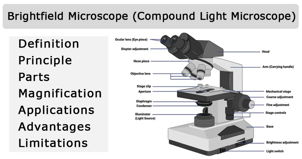
Brightfield Microscope (Compound Light Microscope ...

Labeling the Parts of the Microscope | Microscope activity ...
Label microscope - Teaching resources
Parts of a Microscope

Labeling a Microscope Free Worksheet Pack

Parts of a Microscope - SmartSchool Systems
Parts Of A Microscope Labeling Teaching Resources | TpT

Microscope labeling

Simple Microscope - Diagram (Parts labelled), Principle ...

Free Microscope Drawing, Download Free Microscope Drawing png ...

This is a common compound microscope. Label its parts from A ...

Label the Microscope Diagram | Download Scientific Diagram
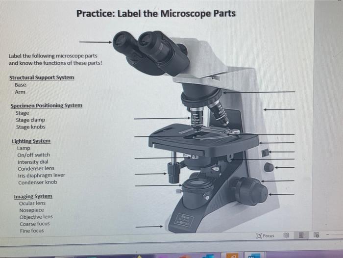
Solved Practice: Label the Microscope Parts Label the | Chegg.com

Parts of Stereo Microscope (Dissecting microscope) – labeled ...

Compound Microscope Parts, Function, & Diagram | What is a ...

The Parts of a Microscope (Labeled) Printable Printable (6th ...
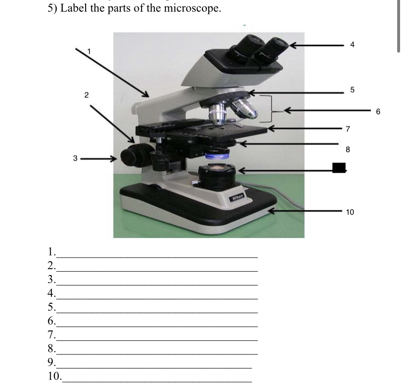
Answered: 5) Label the parts of the microscope. 1… | bartleby

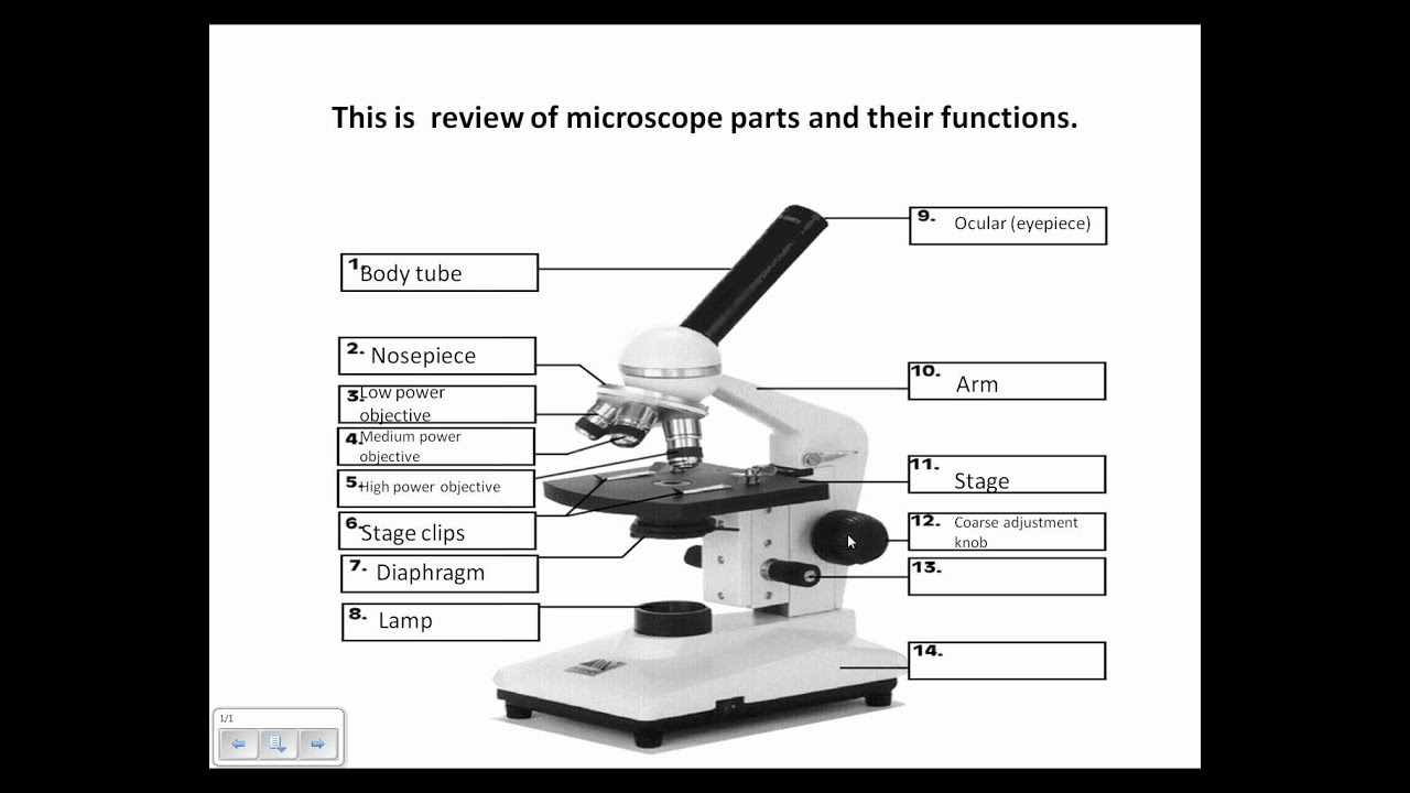
Label the numbered parts of the microscope - ppt download
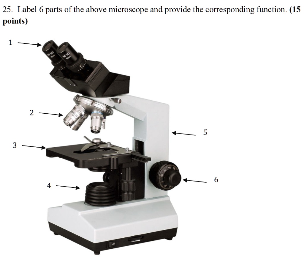
SOLVED:25. Label 6 parts of the above microscope and provide ...
Microscope Review.wmv - YouTube

How to draw and label the parts of a microscope? What are at ...

ᐅ Microscope Construction Simply Explained ᐅ Label ...

Microscope Parts Review Diagram | Quizlet

Microscope Parts (Week 2- Complete).docx - 1 Name: _ Date: _ ...

Microscope Diagram Labeled, Unlabeled and Blank | Parts of a ...

Compound Microscope- Definition, Labeled Diagram, Principle ...

PARTS OF MICROSCOPE| LEARN TO LABEL COMPOUND MICROSCOPE| JUST ...

The Parts of a Compound Microscope and How To Handle Them ...
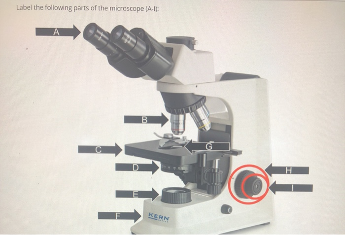
Solved Label the following parts of the microscope (A-1 ...

Simple Microscope - Diagram (Parts labelled), Principle ...
Label a microscope - Teaching resources
Compound and Stereo- microscopes - Microscopes 4 Schools

Microscope Labeling Game

List: Parts of a Microscope and their Function | Pathwooded
Microscope Parts & Specifications | Microscope World Resources
Post a Comment for "40 label the different parts of the microscope"