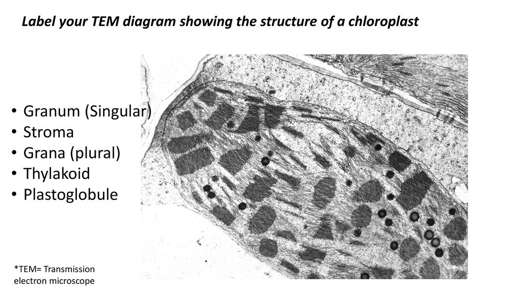
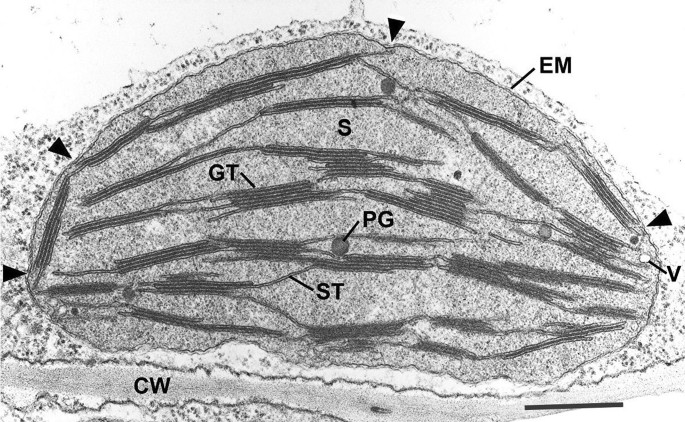
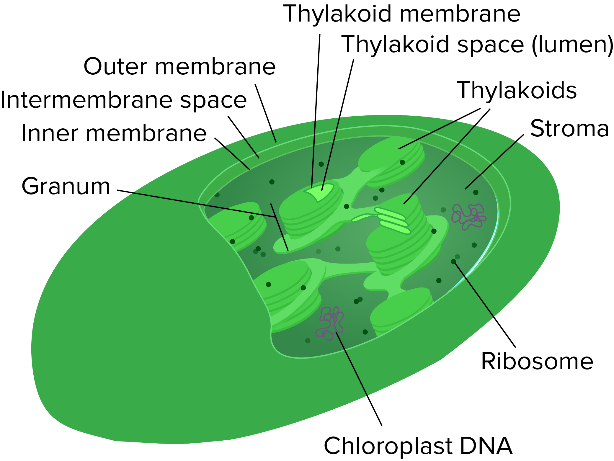
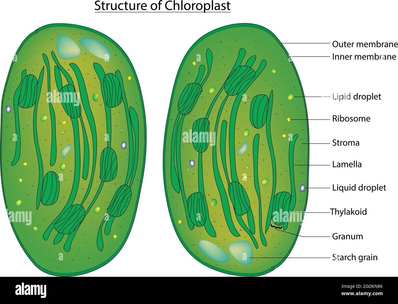
39 transmission electron microscope image of chloroplast labeled
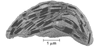
Transmission electron microscopic images of chloroplasts ... Transmission electron microscopic images of chloroplasts and mitochondria in 15-day-old leaves from PRORP1 RNAi mutants and wild-type plants. Transmission electron microscopic images of chloroplasts in ... Download scientific diagram | Transmission electron microscopic images of chloroplasts in WT and OsCpn60β1 mutants. (A) The cell of WT; (B) the cell of β1-1 ...
Transmission electron microscopic images of chloroplasts and ... 19 Mar 2015 — Transmission electron microscopic images of chloroplasts and mitochondria in 15-day-old leaves from PRORP1 RNAi mutants and wild-type plants.

Transmission electron microscope image of chloroplast labeled
Transmission electron microscopic images of chloroplasts ... Transmission electron microscopic images of chloroplasts in mesophyll cells of 2-week-old seedlings of Jimai5265 and Jimai5265yg. a Overview of chloroplasts ... Scanning Electron Microscopic Study of Modified Chloroplasts ... by H Terai · 2000 · Cited by 14 — Scanning electron micrographs of various stages of chloroplasts in the sepal cells of broccoli florets during storage at 20 °C. In the preparation of specimens. Transmission electron microscopy (TEM) images of ... Download scientific diagram | Transmission electron microscopy (TEM) images of chloroplasts from the primary leaf of control (a), NO2-treated (b-d) and ...
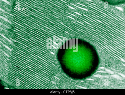
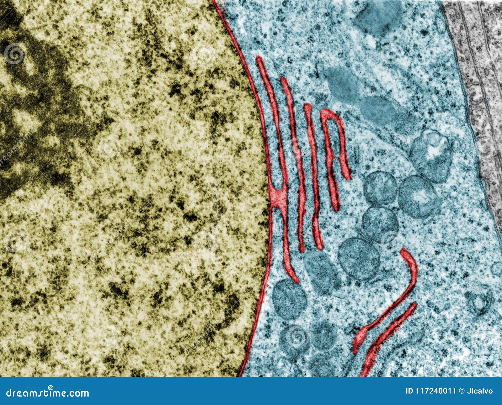
Transmission electron microscope image of chloroplast labeled. Electron microscopy images of chloroplasts from wild type (WT ... Download scientific diagram | Electron microscopy images of chloroplasts from wild type (WT) and the RNAi-W1-7 plants (W1-7). Leaf segments (2 × 2 mm) from ... A brief history of how microscopic studies led to the ... by LA Staehelin · 2020 · Cited by 15 — This review is organized into three chronological sections: During the classic light microscope period (1678–1940), the development of improved ... Transmission electron microscopy picture of the ... Transmission electron microscopy picture of the chloroplast envelopes. Highly magnified view of the outer area of a chloroplast displaying contact sites ... Transmission electron microscopy (TEM) images of ... Download scientific diagram | Transmission electron microscopy (TEM) images of chloroplasts from the primary leaf of control (a), NO2-treated (b-d) and ...
Scanning Electron Microscopic Study of Modified Chloroplasts ... by H Terai · 2000 · Cited by 14 — Scanning electron micrographs of various stages of chloroplasts in the sepal cells of broccoli florets during storage at 20 °C. In the preparation of specimens. Transmission electron microscopic images of chloroplasts ... Transmission electron microscopic images of chloroplasts in mesophyll cells of 2-week-old seedlings of Jimai5265 and Jimai5265yg. a Overview of chloroplasts ...
Transmission electron microscopy (TEM) images of chloroplasts ...
Chloroplast hi-res stock photography and images - Alamy

Warm Up 11/4/13 | Heena Bio HL YAY

Transmission electron micrograph showing the immunogold ...
What is a diagram of a plant and animal cell under an ...

Transmission Electron Micrograph Of A Chloroplast High-Res ...

Cell Micrographs | BioNinja

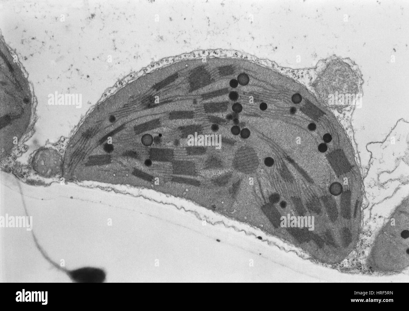
Transmission electron microscopy images of the cross-sections ...

Electron Microscopy of Plant Cells (4.2) | Edexcel ...

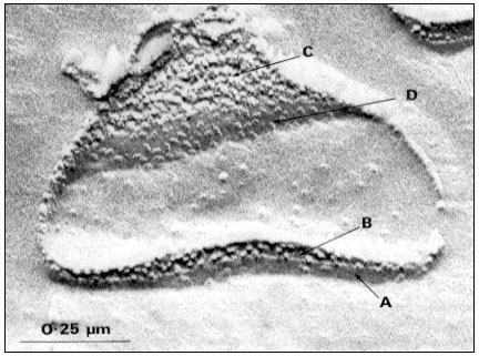
What are the labels of the transmission electronic microscope ...

Draw a labelled diagram of a plant cell as revealed by an electron microscope. | 9 | IMPROVEMEN...
Native architecture of the Chlamydomonas chloroplast revealed ...
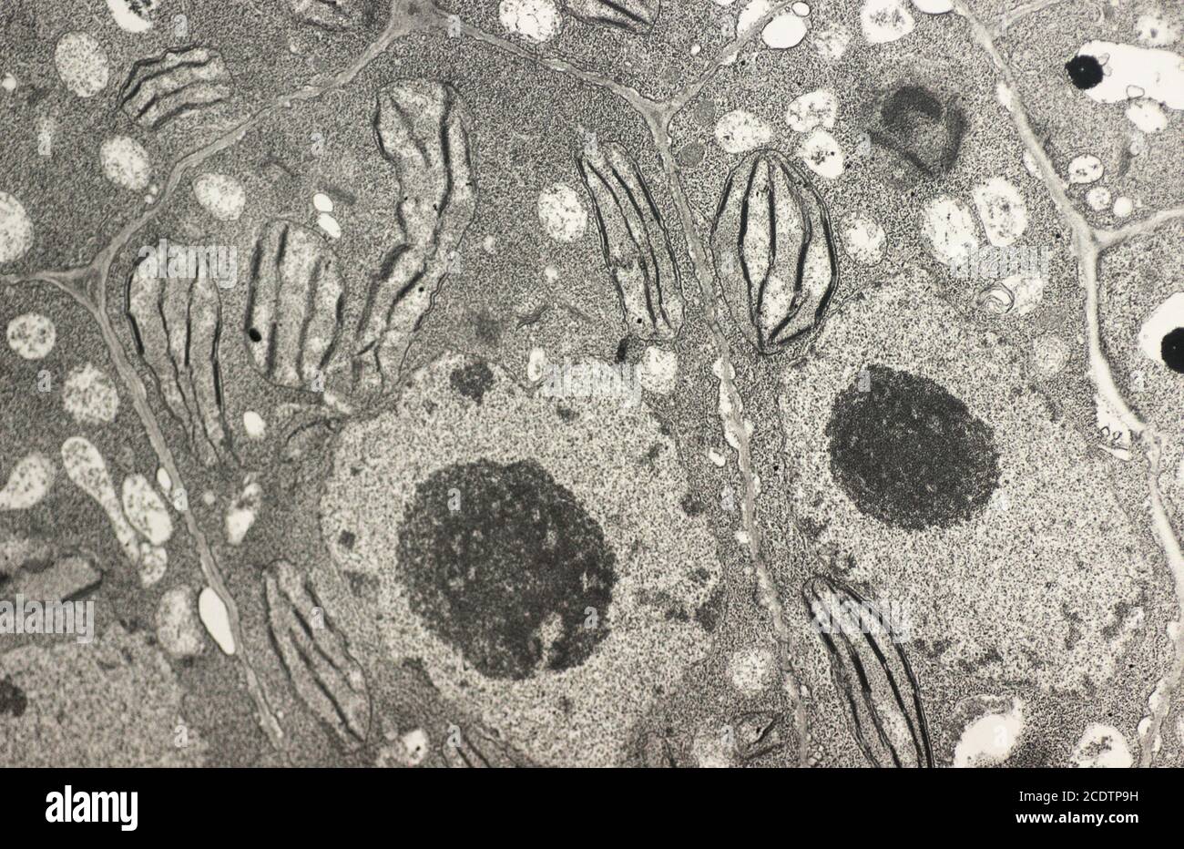
Tem chloroplast hi-res stock photography and images - Alamy
DUE TODAY: SYLLABUS/HONOR CODE SIGNED - ppt download

Cell theory, Plant cell diagram, Cell diagram

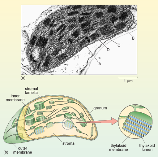
Transmission Electron Microscopy of Chloroplasts and ...
A brief history of how microscopic studies led to the ...
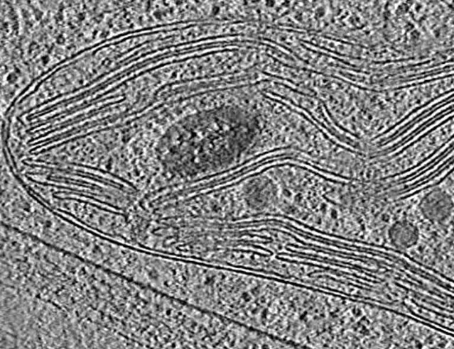
Thylakoids hi-res stock photography and images - Alamy
Tem chloroplast hi-res stock photography and images - Alamy

Thin section electron micrograph of a young tobacco ...
A tour of the cell: View as single page
Nuclear envelope. TEM stock image. Image of micrograph ...

TEM of chloroplast from Coleus blumei - Stock Image - B110 ...

Electron micrograph of an Arabidopsis leaf chloroplast ...

Cell Micrographs | BioNinja

Cell Micrographs | BioNinja

Chloroplast
Mitochondria and chloroplasts (article) | Khan Academy
Scanning Electron Microscopic Study of Modified Chloroplasts ...

Thin section electron micrograph of a young tobacco ...
A) Electron micrograph of a chloroplast of Sphaerosporoceros ...

Transmission electron microscopy (TEM) analysis of ...

Transmission electron microscopy (TEM) analysis of leaf ...

Draw a labelled diagram of chloroplast as seen under an ...
The Molecular Biology of Plant Cells

plant Various Images - SEM | Electron microscope images ...
Tem chloroplast hi-res stock photography and images - Alamy
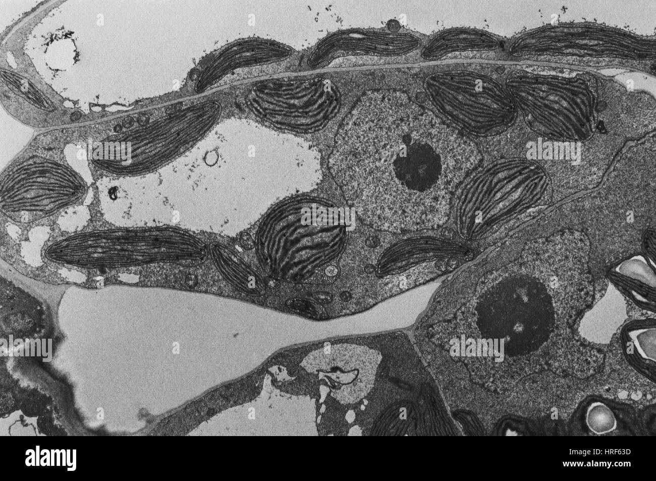
Chloroplast structure hi-res stock photography and images - Alamy
chloroplast | PMG Biology
Post a Comment for "39 transmission electron microscope image of chloroplast labeled"