45 label the transmission electron micrograph of the nucleus
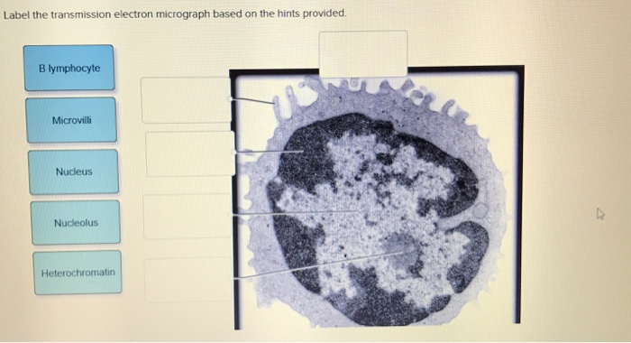
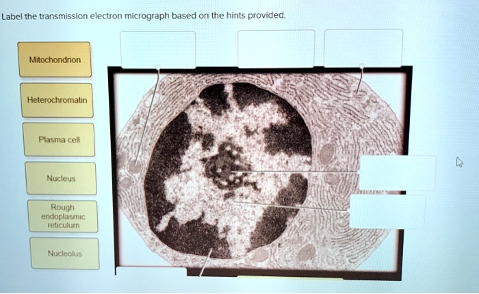
Transmission electron microscopy of the cell nucleus Transmission electron microscopy of the nucleus: This image is an electron spectroscopic imaging (also known as energy filtered transmission electron microscopy) image of a mouse 10T1/2 fibroblast nucleus. Quantitative distributions of phosphorus, which highlights DNA and RNA due to its inherent high phosphorus content were false-coloured green. A&P Unit 2 Exam Flashcards | Quizlet Label the transmission electron micrograph based on the hints provided. Plasma cells produce antibody molecules. Name the cells included in the mononuclear phagocytic system. macrophages monocytes neutrophils An immunoglobulin molecule is an antigen secreted by T lymphocytes. False trabecula of spleen thoracic duct Disease-causing agents are called
Bio101 - Ch 6 HW Flashcards | Quizlet -transmission electron microscopy (TEM) to study the movement of organelles within a living cell -scanning electron microscopy (SEM) to study the detailed movements of living cells -cell fractionation to study the function of specific organelles Beginning within the nucleus, the first step leading to the synthesis of a polypeptide is __________.
Label the transmission electron micrograph of the nucleus
Label This Transmission Electron Micrograph : The Corresponds To The ... Label the transmission electron micrograph of the nucleus. Transmission electron microscopy (tem) is a microscopy technique in which a beam of electrons is transmitted through a specimen to form an image. Subset of labeled images and transfer labels to the entire image corpus. Label the transmission electron micrograph of the. Cell Micrographs | BioNinja A micrograph is a photo or digital image taken through a microscope to show a magnified image of a specimen While organelles have identifying structures, specific shapes may vary depending on the location of cross-sections Prokaryotic Cell Features Feature: none nucleoid cell wall pili flagella all Eukaryotic Cell Features e 02 Quiz Label the transmission electron micrograph of the rn Nucleus ... Professional Academic Writers. Out of thousands who apply to work with us, we only accept the top 3% of the writers. We are cautious about our onboarding process, and every writer undergoes a series of academic tests to evaluate their credibility.
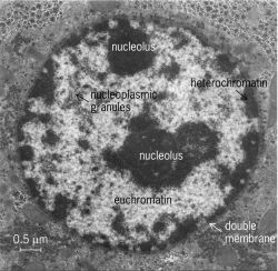
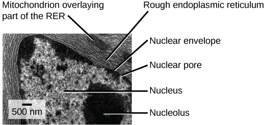
Label the transmission electron micrograph of the nucleus. 8.2: Transmission Electron Microscopy - Chemistry LibreTexts Transmission electron microscopy (TEM) is a form of microscopy which in which a beam of electrons transmits through an extremely thin specimen, and then interacts with the specimen when passing through it. The formation of images in a TEM can be explained by an optical electron beam diagram in Figure 8.2.1. Solved Label the transmission electron micrograph based on - Chegg Question: Label the transmission electron micrograph based on the hints provided Mitochondrion Heterochromatin Plasma cell Nucleus Rough endoplasmic reticulum Nucleolus This problem has been solved! You'll get a detailed solution from a subject matter expert that helps you learn core concepts. See Answer Show transcribed image text Expert Answer Triple and double twin interfaces in magnesium—the role of ... Transmission electron microscopy Abstract Twin boundaries have been shown to deviate from the twinning planes in hcp metals, and facets have often been observed in twin interfaces. Nucleus (TEM) | The Cell - Histology Guide Nucleus. The nucleus of a pancreatic β cell (which synthesizes large amounts of the protein insulin) as seen by transmission electron microscopy (TEM). Switch between the grayscale and color images (see above) to identify these components of a nucleus. Chromatin (blue) - DNA bound to histones and other proteins. Exists in two forms:
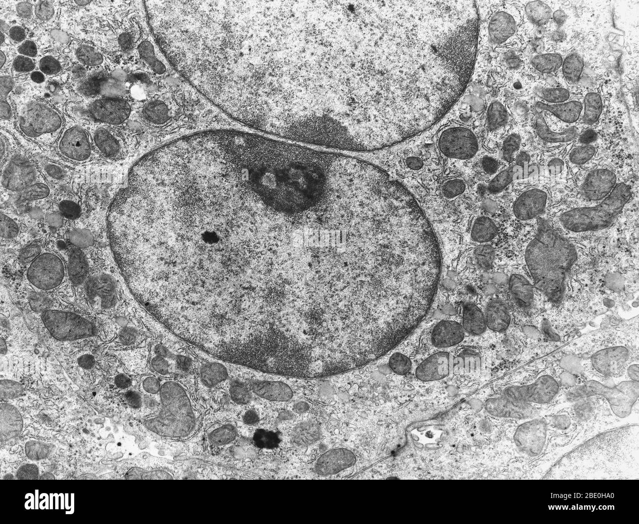
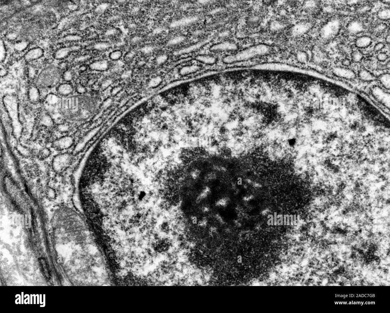
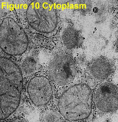
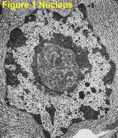
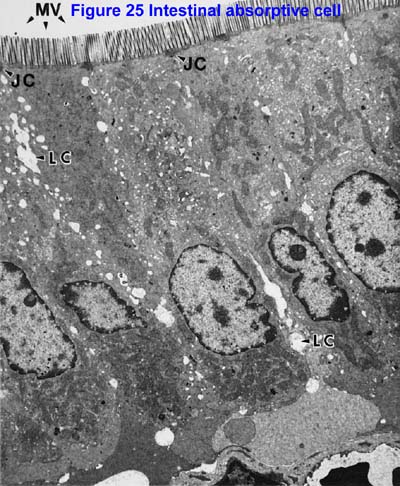
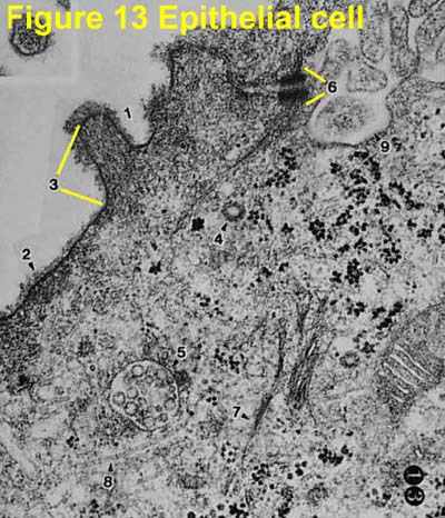
Electron Micrographs - University of Oklahoma Health Sciences Center Below is a collection of electron micrographs with labelled subcellular structures that you should be able to identify. Also, be sure to observe any electron micrographs which are made available in the laboratory by the instructor. You should concentrate on the similarities in form that permit Transmission electron micrograph of cell nucleus - Stock Image - G455 ... Transmission electron micrograph of the nucleus (circular) of a mouse liver cell. Surrounding the nucleus is delicate nuclear membrane, which contains gaps called nuclear pores that allow large molecules to pass out into the cell cytoplasm. The dark area in the lower part of the nucleus is the nucleolus. Anatomy 10.png - Label the transmission electron micrograph... Anatomy 10.png - Label the transmission electron micrograph... Doc Preview Pages 1 Total views 100+ Utah Valley University ZOOL Introduction to Human Anatomy and Physiology BB emileeroylance19 02/01/2020 75% (4) End of preview Upload your study docs or become a member. View full document Become a Member Transmission electron micrograph: nucleus | thank you science Transmission electron micrograph: nucleus. June 24, 2016 Ultimate order, the cell. The nuclear membrane is interesting, to say the least, the perinuclear space the "literal outside" and the two bounding membranes fluid and changing positions in an active and rarely appreciated way. I can remember in the late 1960s working at a place called ...
Microbiology Module 3 Flashcards | Quizlet Please label the image to assess your knowledge of the transport process in eukaryotic cells. nucleus --> RER --> Golgi apparatus --> vesicles --> secretion DNA is copied into RNA in the nucleus, and then the RNA is passed through the nuclear pores to the ribosomes on the rough ER. Labeling the Cell Flashcards | Quizlet Label the transmission electron micrograph of the nucleus. membrane bound organelles golgi apparatus, mitochondrion, lysosome, peroxisome, rough endoplasmic reticulum nonmembrane bound organelles ribosomes, centrosome, proteasomes cytoskeleton includes microfilaments, intermediate filaments, microtubules Identify the highlighted structures Transmission Electron Microscope (With Diagram) - Biology Discussion The final image in a TEM is known as transmission electron micrograph. The salts of some heavy metals, e.g., lead; osmium, tungsten and uranium are often used for staining. These heavy metal stains are used to increase the contrast between ultra structures and the background. The metals can be fixed on to the specimen and is referred to as ... The Cell: The Histology Guide - University of Leeds This picture shows an electron micrograph of a nucleus. The short white arrows are pointing to nuclear pores. Note the appearance of eu- and heterochromatin, and the nucleolus. Heterochromatin stains more densely than euchromatin, but they are both forms of chromatin. Chromatin is the name for the diffuse granular mass of DNA found in ...
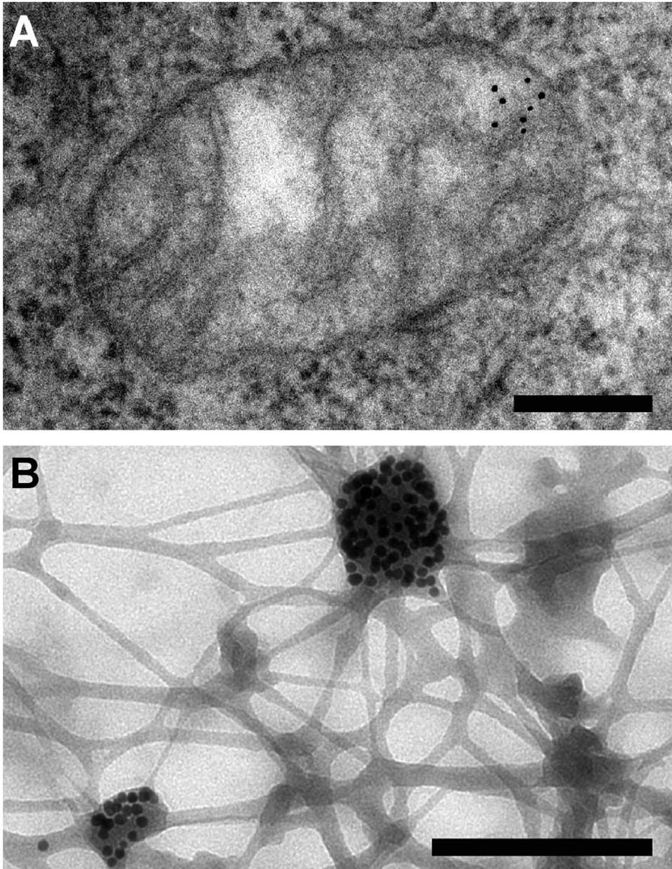
Immunogold labelling - Wikipedia
Neuron under Microscope with Labeled Diagram - AnatomyLearner The nucleus is the spherical or elliptical structure in the neuron containing euchromatic staining (pale staining). Again, the shape of the nucleus of a neuron is generally large because of the little cell body cytoplasm. There is a prominent nucleolus evident in the nucleus of a neuron.
Solved Label the transmission electron micrograph of the ...
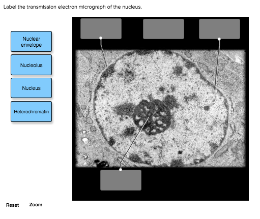
Solved Label the transmission electron micrograph of the - Chegg Question: Label the transmission electron micrograph of the nucleus. Nuclear envelope Nucleolus Nucleus Heterochromatin Reset Zoom Show transcribed image text Expert Answer 100% (25 ratings) Transcribed image text: Label the transmission electron micrograph of the nucleus. Nuclear envelope Nucleolus Nucleus Heterochromatin Reset Zoom

587 Transmission Electron Micrograph Images, Stock Photos ...
The Transmission Electron Microscope | CCBER - UC Santa Barbara Transmission electron microscopes (TEM) are microscopes that use a particle beam of electrons to visualize specimens and generate a highly-magnified image. TEMs can magnify objects up to 2 million times. In order to get a better idea of just how small that is, think of how small a cell is.
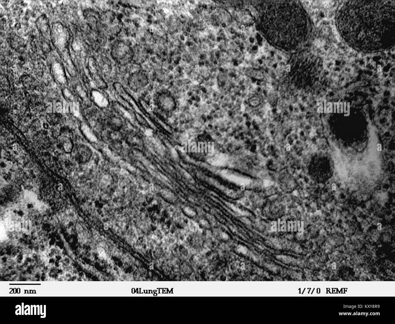
Golgi tem hi-res stock photography and images - Alamy
Label This Transmission Electron Micrograph - Observation on three ... Label the transmission electron micrograph of the nucleus. ImagiNations - Cool Magnified Images from Figures label this transmission electron micrograph ( 16, . Transmission electron microscopy (tem) is a microscopy technique in which a beam of electrons is transmitted through a specimen to form an image.

Labeling the Cell Flashcards | Quizlet
Transmission Electron Microscope (TEM)- Definition, Principle, Images The working principle of the Transmission Electron Microscope (TEM) is similar to the light microscope. The major difference is that light microscopes use light rays to focus and produce an image while the TEM uses a beam of electrons to focus on the specimen, to produce an image. Electrons have a shorter wavelength in comparison to light which ...

TEM of animal cell - Stock Image - G450/0055 - Science Photo ...
Solved Label the transmission electron micrograph of the - Chegg Label the transmission electron micrograph of the cell. 0 Nucleus rences Mitochondrion Heterochromatin Peroxisome Vesicle ULAR bumit Click and drag each label into the correct category to indicate whether it pertains to the cytoplasm or the plasma membrane.

Labeling the Cell Flashcards | Quizlet
Fig. 4: Transmission Electron Micrograph of a Nucleus Fig. 4: Transmission Electron Micrograph of a Nucleus. Transmission electron micrograph of a pancreatic acinar cell from a bat. Note the nuclear membrane, nucleolus, and nuclear pores (arrows). Endoplasmic reticula is also seen.

Transmission electron micrograph of turkey spermatozoa ...
Electron Micrographs of Cell Organelles | Zoology - Biology Discussion The Electron Micrograph of Nucleus: This is an electron micrograph of nucleus. (Fig. 17 & 18): (1) Nucleus was discovered by Brown (1831). (2) It is a characteristic entity of almost all eukaryotic cells except mammalian RBCs. (3) The nucleus is generally one but may also be two, four or many.
Solved FIGURE 3.5 million electron micrograph Celutar | Chegg.com
Label the transmission electron micrograph of the nucleus. - Transtutors Label the transmission electron micrograph of the nucleus. Expert's Answer Solution.pdf Next Previous Related Questions Q: Label this transmission electron micrograph of relaxed sarcomeres by clicking and dragging the... Posted 6 months ago Q: Cells can be represented by a type of model called a cell diagram.
Transmission Electron Micrograph (TEM) of a cell, showing the ...
100 Electron Micrograph Nucleus Premium High Res Photos - Getty Images Find Electron Micrograph Nucleus stock photos and editorial news pictures from Getty Images. Select from premium Electron Micrograph Nucleus of the highest quality. ... carcinoma cell, colored transmission electron micrograph (tem) - electron micrograph nucleus stock illustrations. scanning electron micrograph (sem) of white blood cell ...
The Cell: The Histology Guide
BIO 224: Lab Midterm Review Flashcards | Quizlet Loosely coiled fibers containing protein and DNA within the nucleus. false. T or F: The division of the cell's nuclear parts is called interphase. 46. ... Label the transmission electron micrograph of the mitochondrion. Synthesizes protein for secretion, insertion into the plasma membrane, and lysosomal enzymes. ...

Transmission electron microscopic images of chloroplasts and ...
e 02 Quiz Label the transmission electron micrograph of the rn Nucleus ... Professional Academic Writers. Out of thousands who apply to work with us, we only accept the top 3% of the writers. We are cautious about our onboarding process, and every writer undergoes a series of academic tests to evaluate their credibility.
Electron Micrographs
Cell Micrographs | BioNinja A micrograph is a photo or digital image taken through a microscope to show a magnified image of a specimen While organelles have identifying structures, specific shapes may vary depending on the location of cross-sections Prokaryotic Cell Features Feature: none nucleoid cell wall pili flagella all Eukaryotic Cell Features
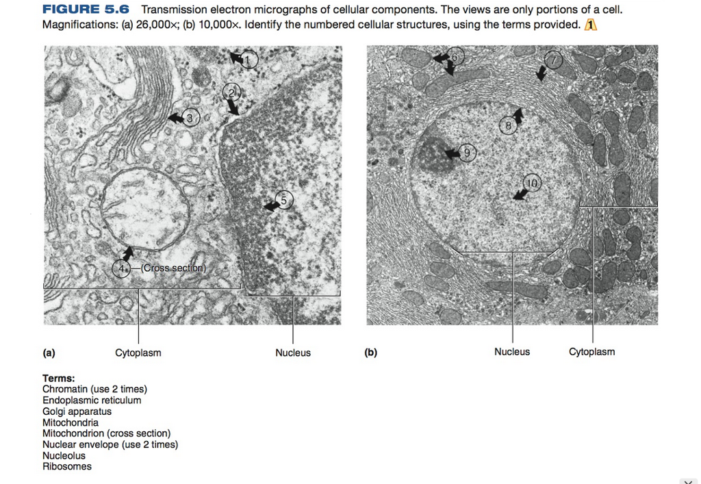
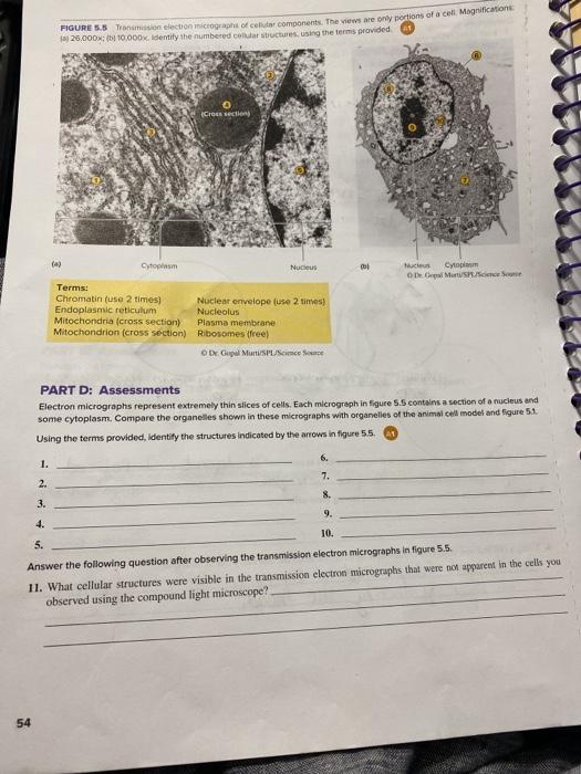
Solved FIGURE 5.6 Transmission electron micrographs of ...
Label This Transmission Electron Micrograph : The Corresponds To The ... Label the transmission electron micrograph of the nucleus. Transmission electron microscopy (tem) is a microscopy technique in which a beam of electrons is transmitted through a specimen to form an image. Subset of labeled images and transfer labels to the entire image corpus. Label the transmission electron micrograph of the.
Solved Label the transmission electron micrograph based on ...

Solved Mitochondrion Nucleus Vesicle Peroxisome | Chegg.com

Nanomaterials | Free Full-Text | A Guide for Using ...

Identifying the Parts of the Nucleus in an Electron Micrograph
Transmission electron micrograph (TEM) showing the nucleus ...
Electron Micrographs
What is a diagram of a plant and animal cell under an ...

Cell Micrographs | BioNinja
Electron Micrographs

Representative transmission electron micrographs of pancreas ...
Electron Micrographs

Cell Micrographs | BioNinja

587 Transmission Electron Micrograph Images, Stock Photos ...

Sub-urothelium. Electron microscopy. (A) A fibroblast (FB ...
SOLVED: Label the transmission electron ricrograph based on ...

Electron micrographs of sperm cell of Ceratopteris richardii ...
Electron Micrographs

Transmission electron microscopy micrographs of labeled MSC ...
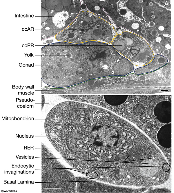
Hermaphrodite Coelomocyte System
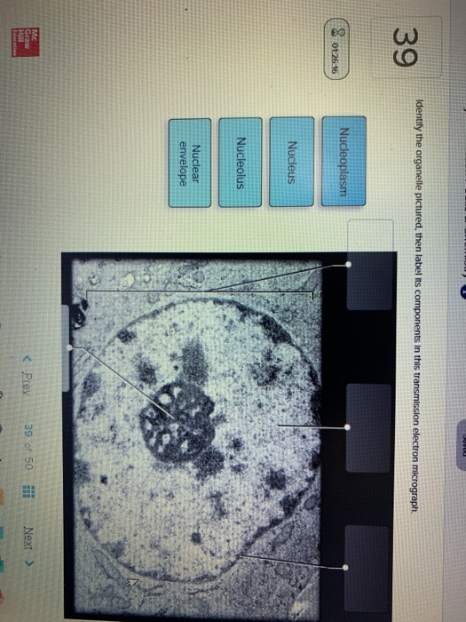
Solved Identify the organelle pictured, then label its ...
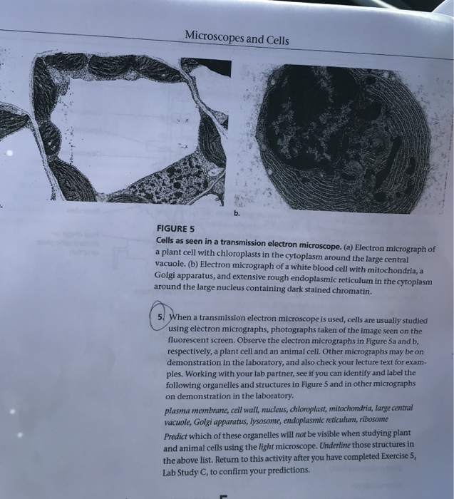
Microscopes and Cells b. FIGURE 5 Cells as seen in a | Chegg.com
Electron Micrographs

Transmission electron microscopy images of SNO cells exposed ...
Organelle and micrograph hi-res stock photography and images ...
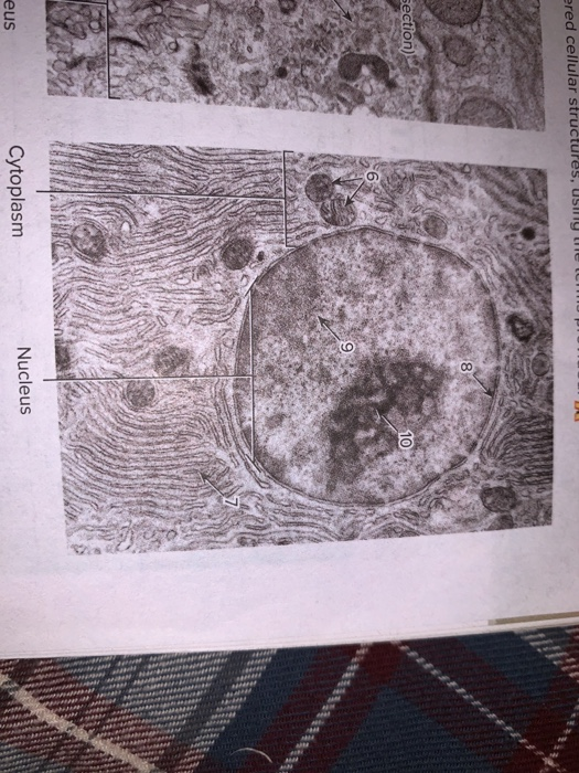
Solved FIGURES Gross section 10 Cytoplasm (a) Nucleus | Chegg.com

Labeling the Cell Flashcards | Quizlet

TEM images of cell organelles such as the nucleus and ...
Cells (2.1, 2.2, 2.3, 2.4 & 2.5) Flashcards | Quizlet

A&P Unit 2 Exam Flashcards | Quizlet
Biology, The Cell, Cell Structure, The Endomembrane System ...

PDF) IB Questionbank Test | Ankit Mistry - Academia.edu
Post a Comment for "45 label the transmission electron micrograph of the nucleus"