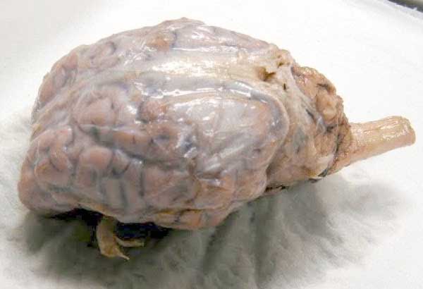
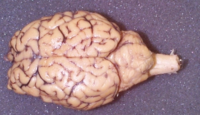
43 unlabeled sheep brain
PDF Lab: Sheep Brain Dissection - Mrs. Moretz's Science Site Sheep brains, although much smaller than human brains, have similar features and can be a valuable addition to anatomy studies. See for yourself what the cerebrum, cerebellum, spinal cord, gray matter, white matter, and other parts of the brain look like! Observation: External Anatomy 1. You'll need a preserved sheep brain for the dissection. Sheep Brain Dissection Guide - The Biology Corner The sheep brain is quite similar to the human brain except for proportion. The sheep has a smaller cerebrum. Also the sheep brain is oriented anterior to posterior whereas the human brain is superior to inferior. 1. The tough outer covering of the sheep brain is the dura mater, one of three meninges (membranes) that cover the brain.
Sheep Brain Label - The Biology Corner A drawing of the brain with the parts unlabeled. Students can practice naming the parts of the brain, then check their answers with the provided key. ... _____ Label theBrain of the Sheep. Publisher: Biologycorner.com; follow on Google+ This work is licensed under a Creative Commons Attribution-NonCommercial 3.0 Unported License. Brain Label ...
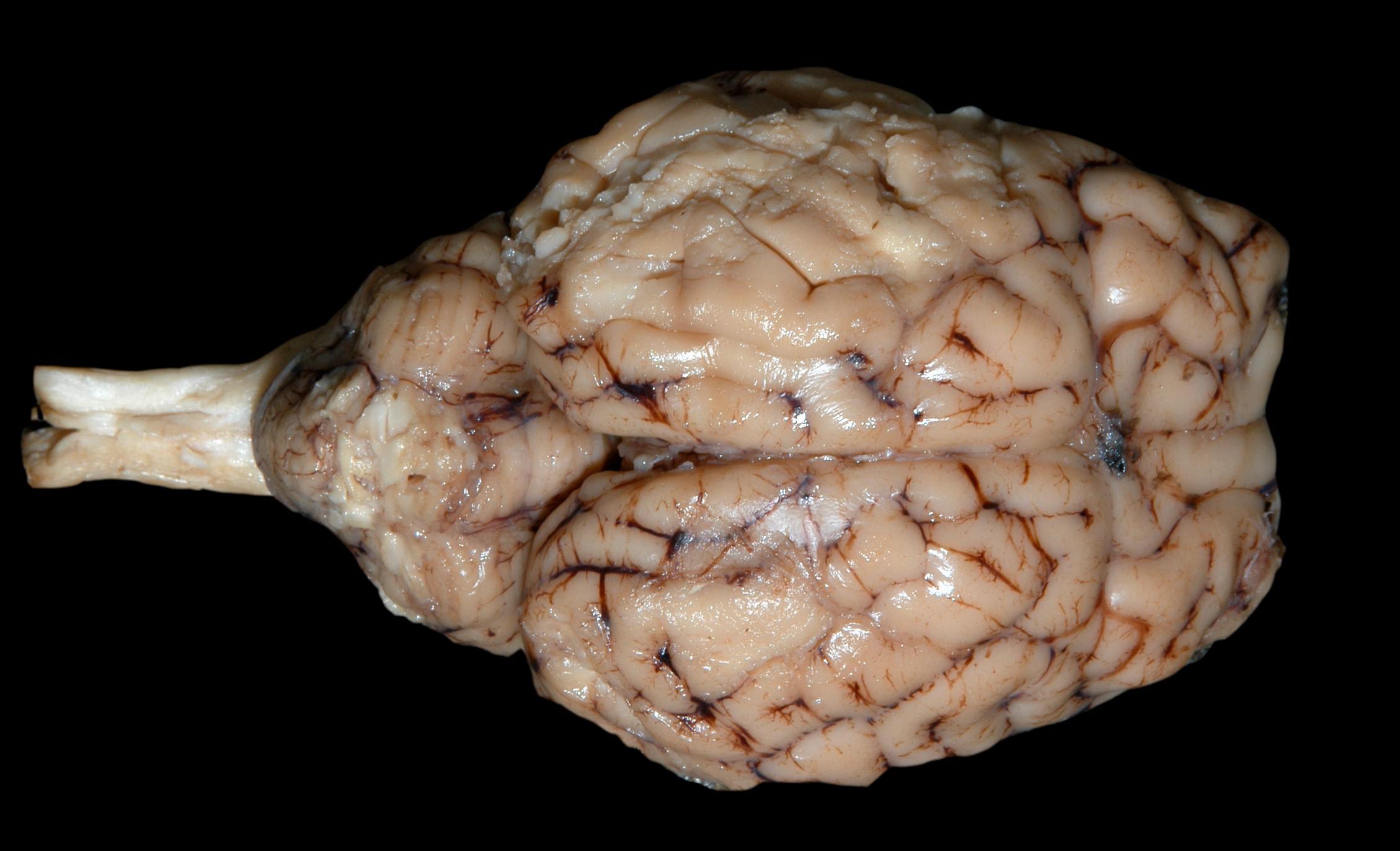
Unlabeled sheep brain
Sheep brain Flashcards | Quizlet Identification of structures observed during sheep brain dissection. Terms in this set (29) dura mater Identify the covering. cerebrum Identify the major brain region. cerebellum Identify the major brain region. olfactory bulb Identify the tip. optic nerve Identify the nerve by name. optic chiasma Identify the "x". optic chiasma 11.7: Sheep Brain Dissection - Biology LibreTexts Background Information: The sheep brain is remarkably similar to the human brain. One major difference, however, is in proportion. For example, the sheep brain has a proportionately smaller cerebrum. Another difference is in orientation of the spinal cord. The sheep spinal cord is orientated anterior to posterior, as in any four-legged animal. Sheep brain unlabeled - YouTube Nov 4, 2013 ... Sheep brain unlabeled. Sam Hirt. Sam Hirt. 528 subscribers. Subscribe. 0. I like this. I dislike this. Share. Share. Save. Save.
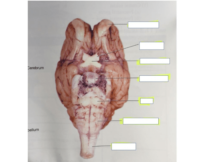
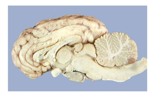
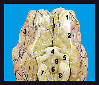
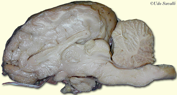
Unlabeled sheep brain. BIO201-Sheep Brain - Udo Savalli Identify the listed structures on the preserved sheep brain. Sheep Brain external view unlabeled. Sheep Brain ventral view unlabeled. Sheep Brain internal ... Parts of the brain: Learn with diagrams and quizzes | Kenhub Labeled brain diagram. First up, have a look at the labeled brain structures on the image below. Try to memorize the name and location of each structure, then proceed to test yourself with the blank brain diagram provided below. Labeled diagram showing the main parts of the brain. Sheep brain anatomy quiz Flashcards | Quizlet medulla oblongata sheep brain autonomic functions; controls vomit, cough, sneeze, swallow, suckle reflexes spinal cord sheep brain bundle of nerve fibers that carry messages to and from the brain parietal lobe of the cerebral hemisphere sheep brain integrating sensory information auditory, visual, touch; contains primary somatosensory cortex Sheep Brain - midsagittal Flashcards | Quizlet frontal lobe Identify the lobe labeled 1 parietal lobe Identify the lobe labeled 2 occipital lobe Identify the lobe labeled 3 arbor vitae Identify the structure labeled 4 spinal cord Identify the structure labeled 5 medulla oblongata Identify the structure labeled 6 fourth ventricle Identify the space labeled 7 pons Identify the structure labeled 8
BIO201-Sheep Brain - Savalli This page last updated 18 August 2019 by Udo M. Savalli (dr.udo @ savalli.us)Images and text © Udo M. Savalli. All rights reserved. Sheep Brain Dissection with Labeled Images - The Biology Corner 1. The sheep brain is enclosed in a tough outer covering called the dura mater. You can still see some structures on the brain before you remove the dura mater. Take special note of the pituitary gland and the optic chiasma. These two structures will likely be pulled off when you remove the dura mater. Brain with Dura Mater Intact Sheep brain images | Lab - Amherst College Sheep brain images by Mark Yarchoan '07. First image in each group of 3: plain photo. Second image: with arrows pointing to structures. Lab 9—Sheep Brain—Labeled - Bluegrass Community and Technical College Lab 9—Sheep Brain—Labeled BIO 137 Virtual Lab 9 The Sheep's Brain Return to: The Unlabeled BrainsLab 9 PageBIO 137 Main Page Be sure to practice identifying the structures using the unlabeled photos. This page created and maintained by Udo M. Savalli. Last updated August 13, 2005.
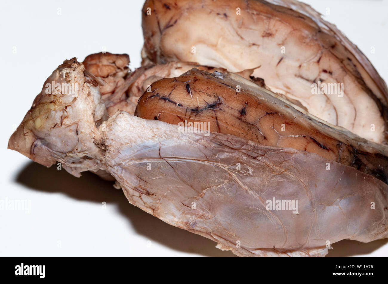
sheep brain labeled Diagram | Quizlet Start studying sheep brain labeled. Learn vocabulary, terms, and more with flashcards, games, and other study tools. Sheep Brain Dissection labeled Diagram | Quizlet Sheep Brain Dissection labeled Diagram | Quizlet Sheep Brain Dissection labeled + − Learn Test Match Created by AllieKlinger Terms in this set (8) Corpus Collosum ... Lateral Ventricle ... Fornix ... Hypothalamus ... Cerebral Aqueduct ... Central Canal ... Inferior Collicuious ... Transverse Fissure ... sydneypfleiger Sheep Brain Dissection Project Guide | HST Learning Center Sheep Brain Dissection: Internal Anatomy Place the brain with the curved top side of the cerebrum facing up. Use a scalpel (or sharp, thin knife) to slice through the brain along the center line, starting at the cerebrum and going down through the cerebellum, spinal cord, medulla, and pons. Sheep Brain Anatomy with Labeled Diagram The sheep brain anatomy consists of 3 major parts - prosencephalon (forebrain), mesencephalon (midbrain), and rhombencephalon (hindbrain). These 3 main parts of the sheep brain again divide into specific segments. There are also 5 different lobes in the sheep brain structure - frontal, parietal, occipital, temporal, and limbic area.
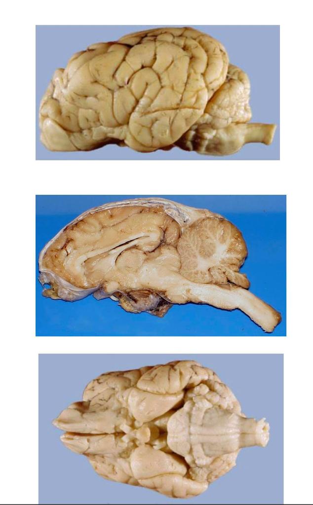
Sheep brain images | Lab | Amherst College
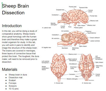
Sheep Brain Dissection | Carolina.com Sheep Brain Dissection The body consists of organ systems, each with specific roles for maintaining life. Each organ system consists of organs that operate together to carry out certain body functions. Organ systems do not operate independently but are coordinated for the organism to survive.

Sheep Brain
Sheep Brain Neuroanatomy Online Self-Test | KPU.ca - Kwantlen ... Sheep Brain Neuroanatomy Online Self-Test Use each diagram as a reference, and selected the correct answer for each lettered structure. You may find it useful to open the diagrams in a separate window to review while answering each question. Dorsal Surface Dorsal Surface A * Occipital Lobe Temporal Lobe Cerebellum Parietal Lobe Dorsal Surface B *
Sheep Brain Practical
Sheep Brain Instructions - University of Scranton The Sheep Brain Dissection Guide Uses Frames And It May Not Be Compatible With All Web Browsers!! You have several options when proceeding through this guide. As you progress, you will see the image of the brain being dissected in this window. ... This button allows you to toggle between labeled and unlabeled images. On many of the images you ...
Brain Dissection Guide
NERVOUS SYSTEM - SHEEP BRAIN IMAGES - sdmesa.edu Sheep Brain Unlabeled. Sheep Brain Leader-Lined. Sheep Brain Labeled. San Diego Mesa College 7250 Mesa College Drive San Diego, CA 92111-4998 Student Support San Diego Community College District San Diego City College San Diego Mesa College San Diego Miramar College San Diego Continuing Education.

Sheep Brain
Sheep Brain - Anatomy Corner ... dissection of the sheep's brain, some structures have been labeled. superior colliculus labeled · brain, lateral view · labeled brain · unlabeled brain ...
Inferior view of sheep brain - 11.11 Quiz
Neuroanatomy: Dissection of the Sheep Brain (This can be a little tricky to find on the sheep brain – let me know if you need help.) Anterior to the central sulcus is the primary motor cortex, posterior ...

Sheep Brain Images
PDF Sheep Brain Practical Study Guide - auburn.k12.il.us Sheep Brain Practical Study Guide. Dura Mater. Olfactory Bulb Pituitary Gland Dura Mater Optic Chiasm. Corpus Callosum Longitudinal Fissure Lateral Ventricle Gray Matter White Matter. Arbor Vitae "Tree of Life" Cerebellum "Little Brain" ...

Physiological Psychology
Sheep brain unlabeled - YouTube Nov 4, 2013 ... Sheep brain unlabeled. Sam Hirt. Sam Hirt. 528 subscribers. Subscribe. 0. I like this. I dislike this. Share. Share. Save. Save.
Sheep Brain Explora on Guide
11.7: Sheep Brain Dissection - Biology LibreTexts Background Information: The sheep brain is remarkably similar to the human brain. One major difference, however, is in proportion. For example, the sheep brain has a proportionately smaller cerebrum. Another difference is in orientation of the spinal cord. The sheep spinal cord is orientated anterior to posterior, as in any four-legged animal.
Solved Structures To Label In The Sheep Brain Whole (uncut ...
Sheep brain Flashcards | Quizlet Identification of structures observed during sheep brain dissection. Terms in this set (29) dura mater Identify the covering. cerebrum Identify the major brain region. cerebellum Identify the major brain region. olfactory bulb Identify the tip. optic nerve Identify the nerve by name. optic chiasma Identify the "x". optic chiasma

Sheep brain Diagram | Quizlet

Sheep Brain
Sheep Brain Dissection with Labeled Images

Sheep Brain Images
Sheep Brain Dissection - Mrs N. Nelson's Science Website
Neuroanatomy: Dissection of the Sheep Brain

Sheep Brain
Dissection of the Sheep Brain
PhysioEx Exercise 3: Neurophysiology
Brain 1a
Sheep brain hi-res stock photography and images - Alamy

Teaching Labs - NeuroscienceCourses.com
Sheep Brain Explora on Guide

Sheep Brain Images

Sheep Brain Flashcards | Quizlet
Lab: Sheep Brain Dissection
Solved Identify and label the highlighted structures below ...

Sheep brain | Atlas of Comparative Vertebrate Anatomy

Sheep Brain Dissection - Behavioral Neuroscience Lab Log

Welcome to the sheep brain structure review! arachnoid layer ...

Physiological Psychology
Sheep Brain Neuroanatomy Online Self-Test | KPU.ca - Kwantlen ...
Lab: Sheep Brain Dissection
Welcome to the sheep brain structure review! arachnoid layer ...
Sheep Brain Dissection Lab
BIO201-Sheep Brain

BIO201-Sheep Brain
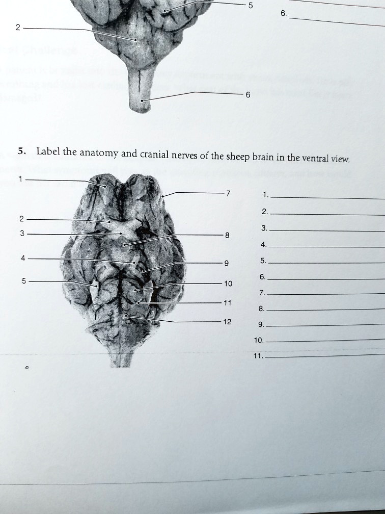
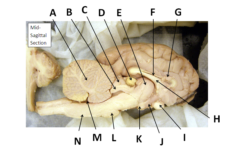
SOLVED: Label the anatomy and cranial nerves of the sheep ...
Sheep Brain Dissection

Sheep Brain Anatomy Quiz #SwagYOLO

Exif | Dorsal Sheep Brain 1 (unlabeled) | Flickr - Photo Sharing!

Sheep Brain Images

Sheep Brain Images
Post a Comment for "43 unlabeled sheep brain"