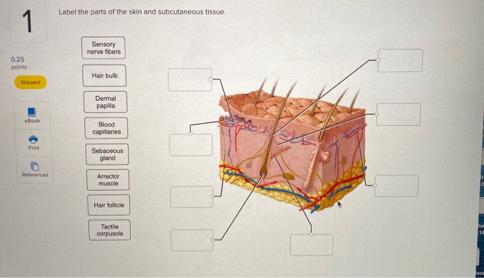
44 label the parts of the skin and subcutaneous tissue
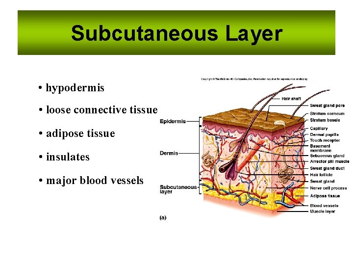
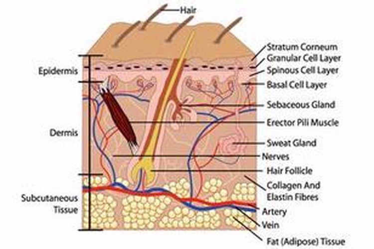
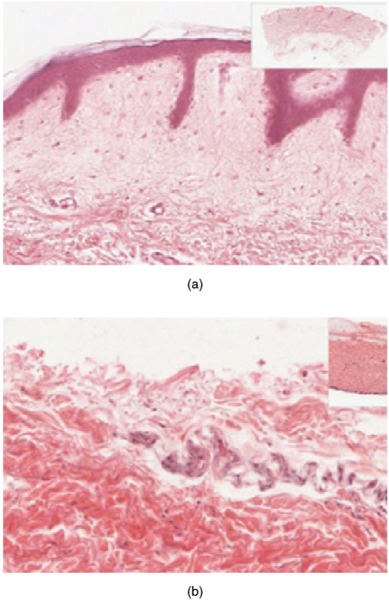
Function And Structure of Skin And Subcutaneous Tissue The epidermis is the thin outer layer of skin, the dermis is the thicker inner layer of skin. Beneath the dermis, lies a layer of loose connective tissue called subcutaneous tissue or the hypodermis deeper tissues including muscles, tendon, ligament, joint capsule and bone lie beneath the subcutaneous tissue layer. Fat removal procedures - Wikipedia The degree of exposure to cooling causes cell death of subcutaneous fat tissue, without apparent damage to the overlying skin. It appears primarily applicable to limited discrete fat bulges. Adverse effects include transient local redness, bruising and numbness of the skin, and these are expected to subside.
PharmaCircle This website uses cookies to help provide you with the best possible online experience. Please read our Terms & Conditions and Privacy Policy for information about ...

Label the parts of the skin and subcutaneous tissue
Solved Label the parts of the skin and subcutaneous tissue. | Chegg.com Question: Label the parts of the skin and subcutaneous tissue. Sensory nerve fibers Hair bulb Piloerector muscle Hair follicle Sebaceous gland Tactile corpuscle Blood capillaries Dermal papilla < Prey, 8 of 50 Next > This problem has been solved! See the answer Show transcribed image text Expert Answer Skin Anatomy - EnchantedLearning.com hair shaft - The part of the hair that is above the skin. ... (sweat). The gland is located in the epidermis; it releases sweat onto the skin. subcutaneous tissue - fatty tissue located under the dermis. Worksheet to Print ... soft, hot, cold, rough, smooth, grainy, sharp. Skin Anatomy Diagram to Label Label the skin anatomy diagram. Or go to ... Tag: label the parts of the skin and subcutaneous tissue Homepage / label the parts of the skin and subcutaneous tissue. Tag: label the parts of the skin and subcutaneous tissue. Label the Diagram of The Skin. By Admin Posted on August 10, 2022. Skin is the largest organ of our body. Together with hair, nails, body glands, and nerves, the structure of the human skin forms the integumentary system ...
Label the parts of the skin and subcutaneous tissue. AHCDWeek2SOL1.pdf - 1. Award: 10.00 points Problems? Adjust... Label the parts of the skin and subcutaneous tissue. Explanation: The skin consists of two layers: a stratified squamous epithelium called the epidermis and a deeper connective tissue layer called the dermis. Below the dermis is another connective tissue layer, the hypodermis, which is not part of the skin. The skin is equipped with a Dupixent (Dupilumab) Subcutaneous: Uses, Side Effects, Dosage Jul 22, 2022 · Wash your hands and clean the area of skin you plan to inject with an alcohol swab. Allow your skin to dry without blowing on it or touching it again. Remove the needle cap with the needle pointing away from yourself. Pinch a section of your skin between your thumb and other fingers, and insert the needle completely at about a 45-degree angle. Impaired Skin Integrity Nursing Diagnosis and 5 Best Care Plans Mar 14, 2022 · Affected area suspected of impaired skin integrity may be hot and tender to touch. There may be observations of fever. Visible damage to integumentary tissues like the cornea, mucous membranes, subcutaneous skin, etc. The area may be inflamed and cause pain to the patient; Guarding of the affected area, and grimacing on contact with affected area How to Give a Subcutaneous Injection (with Pictures) - wikiHow May 29, 2022 · When gathering your skin, do not gather any muscle tissue. You should be able to feel the difference between the soft upper fat layer and firmer, lower muscle tissue. Subcutaneous medications are not intended for injection into the muscle and, if administered into muscle, may result in bleeding into the muscle tissue.
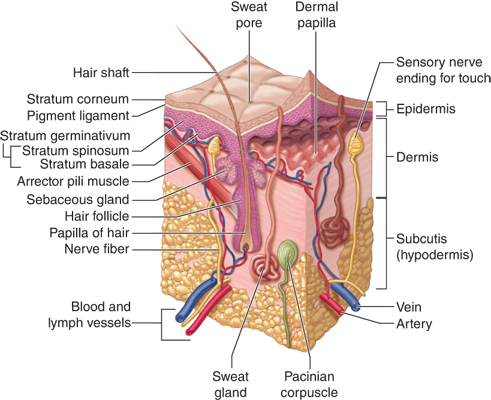
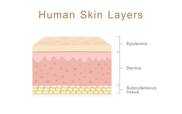
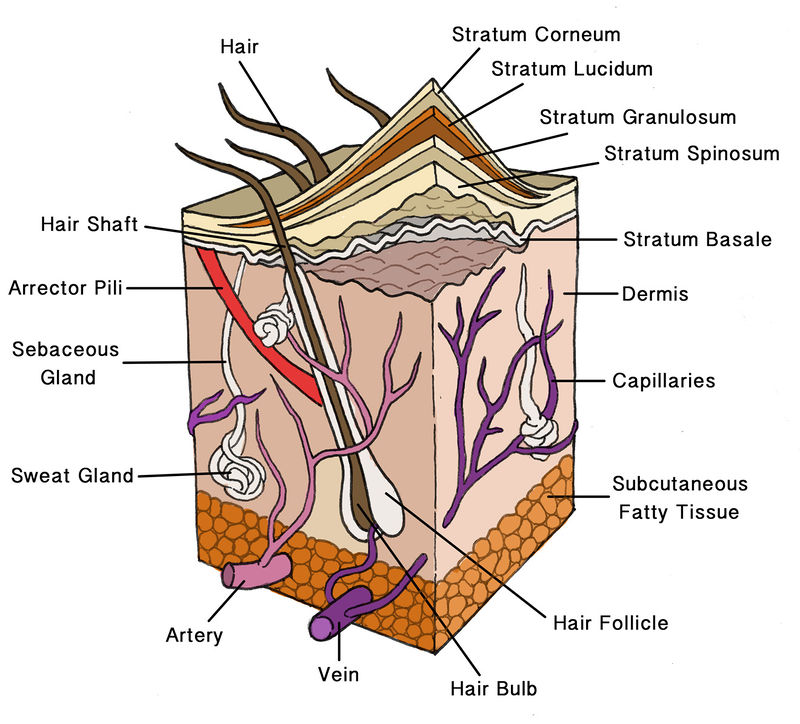
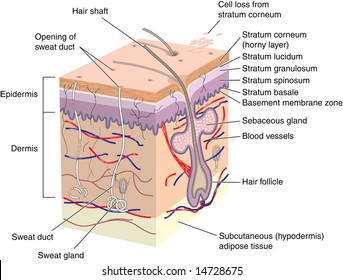
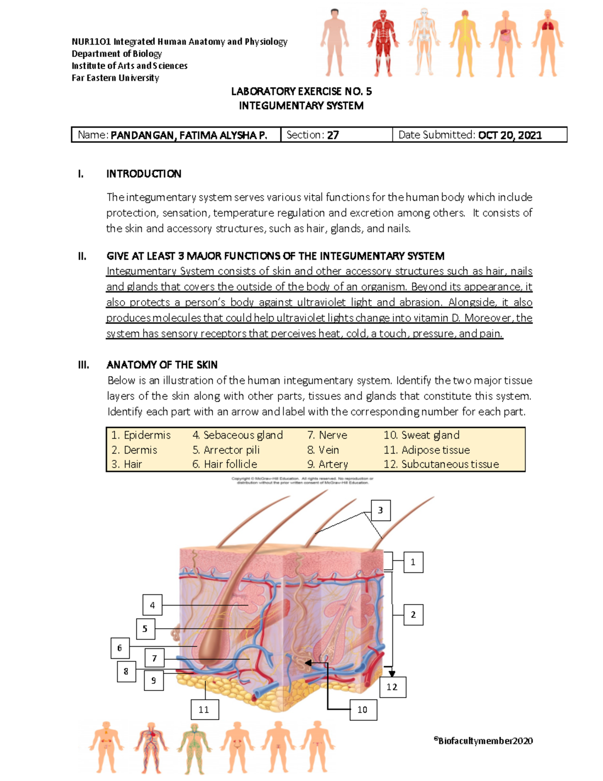
Label the Diagram of The Skin | Portal Sejarah Together with hair, nails, body glands, and nerves, the structure of the human skin forms the integumentary system, which is a system that encloses and protects the inside of the body. Label The Diagram of The Skin 1. Epidermis 2. Dermis 3. Hypodermic 4. Hair shaft 5. Oil glands (sebaceous glands) 6. Sweat glands 7. Blood vessels and nerve endings The Skin: 7 Most Important Layers and Functions - MedicineNet Stratum lucidum. Stratum granulosum. Stratum spinosum. Stratum basale. Dermis. Hypodermis. The first five layers form the epidermis, which is the outermost, thick layer of the skin. All seven layers vary significantly in their anatomy and function. The skin serves various functions that include. The subcutaneous layer: Anatomy, composition, and functions The subcutaneous layer is located underneath the dermis and is one of the three layers of the skin. It is the deepest skin layer, composed of fat cells, collagen, blood vessels, and nerves. The... Hypodermis (Subcutaneous Tissue): Function & Structure - Cleveland Clinic It has many important functions, including storing energy, connecting the dermis layer of your skin to your muscles and bones, insulating your body and protecting your body from harm. As you age, your hypodermis decreases in size, and your skin starts to sag. Dermal fillers help restore volume to your skin as your hypodermis decreases.
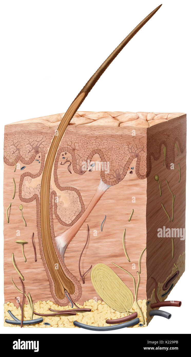
(Get Answer) - Label the parts of the skin and subcutaneous tissue. 0. ... Label the parts of the skin and subcutaneous tissue. 0.22 Cutaneous blood vessels Book Hypodermis Sweat pores Epidermis Hairs Dermis Lamellar corpuscle Sweat gland Draw the structure of pyruvate, showing its appropriate structure at pH 7.4. What does the top pressure gauge in the figure read? Expert's Answer Solution.pdf Next Previous Q: Q: Q: Q: Skin and Subcutaneous Tissue Service Line - thehaugengroup.com In lesson 1, the anatomy and function of structures pertinent to these parts of the body is discussed. This paves the way for discussion of procedures and coding guidelines for procedures performed on the skin, subcutaneous tissue, fascia, and breast in lessons 2 and 3 with special emphasis on excisional and non-excisional debridement, breast ... Layers of Skin: How Many, Diagram, Model, Anatomy, In Order - Healthline Subcutis. The layer of skin beneath the dermis is sometimes called the subcutaneous fat, subcutis, or hypodermis layer. This layer provides insulation for your body, keeping you warm. It also ... Integumentary System – Medical Terminology for Healthcare ... A condition in which conservation of the body core heat results in the skin freezing. Gangrene. Death of tissue due to blood supply loss. Hidradenitis. Inflammation of a sweat gland. Hypodermis . Also known as the subcutaneous layer; the layer of the skin below the dermis that is composed mainly of loose connective and fatty tissues. Incision
Chapter 4: the skin Flashcards | Quizlet
Solved Label the parts of the skin and subcutaneous tissue ... - Chegg Question: Label the parts of the skin and subcutaneous tissue. 0.22 Cutaneous blood vessels Book Hypodermis Sweat pores Epidermis Hairs Dermis Lamellar corpuscle Sweat gland This problem has been solved! See the answer label the parts of the skin and subcutaneous tissue Show transcribed image text Expert Answer 98% (46 ratings) A …
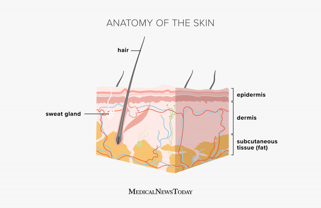
Anatomy and Physiology of the Skin
Understanding The Different Layers Of Skin - SkinKraft Collagen is a fibrous protein that is made of long and thin fibrils that keep the skin cells together. They give strength to the skin. Elastin is a fibrous protein that gives skin its elastic nature. 3. Subcutaneous Tissue. The subcutaneous tissue or hypodermis consists of well-vascularized, loose connective tissue and adipose tissue.

Ch 6 quiz - Integumentary System Flashcards | Quizlet
Anatomy of the Skin | SEER Training - National Cancer Institute Under these two skin layers is a fatty layer of subcutaneous tissue, known as the subcutis or hypodermis. The skin contains many specialized cells and structures: Basket Cells Basket cells surround the base of hair follicles and can sense pressure. They are evaluated when assessing overall nerve health and condition. Blood Vessels

Final Exam A&P 1 Flashcards | Quizlet
Layers of the Skin | Anatomy and Physiology I - Lumen Learning The hypodermis (also called the subcutaneous layer or superficial fascia) is a layer directly below the dermis and serves to connect the skin to the underlying fascia (fibrous tissue) of the bones and muscles. It is not strictly a part of the skin, although the border between the hypodermis and dermis can be difficult to distinguish.
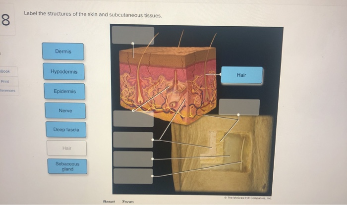
Solved Label the structures of the skin and subcutaneous ...
Subcutaneous Tissue: Composition, Function, Structure - Verywell Health Several structures and specialized cells exist within the subcutaneous tissue. These include: Collagen and elastin fibers (these attach the dermis to muscles and bones) Fat cells Blood vessels Sebaceous glands Nerve endings Hair follicle roots

Part I: Layers of the Epidermis A. The following diagram of ...
Chapter 4: the skin Flashcards | Quizlet The outermost layer of the epidermis is keratinized and known as Stratham. Corneum. The three pigments that contribute to skin color are. Melanin, carotene, and hemoglobin. The sebaceous and sweat glands associated with the skin are classified as ______ glands because they release secretions to the skin surface vie ducts. Exocrine.
The Skin and Subcutaneous Tissue | Basicmedical Key
Skin Structure (Labeling) Flashcards | Quizlet Where is Adipose Tissue Located on the skin structure? Recommended textbook explanations. Introduction to Anatomy and Physiology 1st Edition Michelle Provost-Craig, Susan J. Hall, ... Skin Labeling. 24 terms. Allison_Dolezal. Skull Review. 58 terms. jeffshay. skin layers. 5 terms. Kaeli_Petit. Facial & Cranial Bones. 36 terms. csavier1.

Activity 3. Label and Describe Me! Directions. Label the ...
Skin Cross-Section - Anatomy and Physiology - Innerbody The skin is by far the largest organ of the human body, weighing about 10 pounds (4.5 kg) and measuring about 20 square feet (2 square meters) in surface area. It forms the outer covering for the entire body and protects the internal tissues from the external environment. The skin consists of two distinct layers: the epidermis and the dermis.
Essentials of Human Anatomy Skin 1 Anatomy of
Subcutaneous Tissue Function and What Can Impact Its Health The middle layer of your skin contains sweat glands, lymphatic vessels, blood vessels, connective tissue, and hair follicles. Subcutaneous tissue. The deepest layer of skin is made of connective...

The Skin - Science Quiz
Solved Label the parts of the skin and subcutaneous tissue. | Chegg.com Question: Label the parts of the skin and subcutaneous tissue. Piloerector muscle Tactile corpuscle Dermal papilla Hair follicle Sebaceous gland Blood capillaries Hair bulb This problem has been solved! See the answer Show transcribed image text Expert Answer 100% (13 ratings)

Skin Structure (Labeling) Flashcards | Quizlet
Picture of the Skin - WebMD The dermis, beneath the epidermis, contains tough connective tissue, hair follicles, and sweat glands. The deeper subcutaneous tissue (hypodermis) is made of fat and connective tissue. The skin's...

Skin 1: the structure and functions of the skin | Nursing Times
Chapter 6 Worksheet Flashcards | Quizlet Label the parts of the skin and subcutaneous tissue Label the layers of the skin top to bottom: - stratum corneum - stratum lucidum - stratum granulosum - stratum spinosum - stratum basale - dermis Label the cell types found in the skin Drag each label to the appropriate layer (A, B, or C) for each term or phrase.
Integumentary system parts: Quizzes and diagrams | Kenhub
Anatomy and Physiology Homework Chapter 6 Flashcards | Quizlet The skin is equipped with a variety of nerve endings that react to heat, cold, touch, texture, pressure, vibration, and tissue injury. Label the parts of the skin and subcutaneous tissue. -Hypodermis -Sweat pores -Dermis -Hairs -Cutaneous blood vessels -Epidermis -Sweat gland -Lamellar corpuscle -Hairs -Sweat pores -Cutaneous blood vessels

5.1 Layers of the Skin – Anatomy & Physiology
Skin Anatomy: The Layers of Skin and Their Functions - Verywell Health Subcutaneous tissue is the innermost layer of the skin. It is mostly made up of fat, connective tissues, larger blood vessels, and nerves. 5 The majority of your body fat is stored in the subcutaneous layer. It not only insulates you against changing temperatures but protects your muscles and internal organs from impacts and falls.
21,931 Skin Anatomy Stock Photos, Pictures & Royalty-Free ...
Anatomy, Skin (Integument), Epidermis - StatPearls - NCBI Bookshelf Skin is the largest organ in the body and covers the body's entire external surface. It is made up of three layers, the epidermis, dermis, and the hypodermis, all three of which vary significantly in their anatomy and function. The skin's structure is made up of an intricate network which serves as the body's initial barrier against pathogens, UV light, and chemicals, and mechanical injury ...
Hypodermis hi-res stock photography and images - Alamy
Types of injections: Uses, sites, and what to expect Jul 29, 2021 · Unlike muscle tissue, subcutaneous tissue has few blood vessels, according to a 2017 study. The presence of fewer blood cells allows the body to absorb the medication slowly over a period of time.

340 Subcutaneous Tissue Cliparts, Stock Vector and Royalty ...
Tag: label the parts of the skin and subcutaneous tissue Homepage / label the parts of the skin and subcutaneous tissue. Tag: label the parts of the skin and subcutaneous tissue. Label the Diagram of The Skin. By Admin Posted on August 10, 2022. Skin is the largest organ of our body. Together with hair, nails, body glands, and nerves, the structure of the human skin forms the integumentary system ...

Subcutaneous tissue - Wikipedia
Skin Anatomy - EnchantedLearning.com hair shaft - The part of the hair that is above the skin. ... (sweat). The gland is located in the epidermis; it releases sweat onto the skin. subcutaneous tissue - fatty tissue located under the dermis. Worksheet to Print ... soft, hot, cold, rough, smooth, grainy, sharp. Skin Anatomy Diagram to Label Label the skin anatomy diagram. Or go to ...

The Integumentary System - ppt download
Solved Label the parts of the skin and subcutaneous tissue. | Chegg.com Question: Label the parts of the skin and subcutaneous tissue. Sensory nerve fibers Hair bulb Piloerector muscle Hair follicle Sebaceous gland Tactile corpuscle Blood capillaries Dermal papilla < Prey, 8 of 50 Next > This problem has been solved! See the answer Show transcribed image text Expert Answer
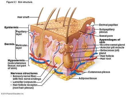
Figure 5-1 The Components of the Integumentary System. - ppt ...
The Integumentary System
Human Biology fig. 1.24 - Layers of the skin - English labels ...
/skin-anatomy-1068880_review-01-9adf9daebac8464eb693274a960bd850-52cb9a92cd394931afe6abfca8074e28.png)
Skin: Anatomy and Function

Reptilian Skin and Its Special Histological Structures ...

Structure and Function of the Skin - Skin Disorders - MSD ...
Subcutaneous tissue hi-res stock photography and images - Alamy

Integumentary System Facts | jpeg integumentary system facts ...

Skin and skin appendage - Knowledge @ AMBOSS

AHCDWeek2SOL1.pdf - 1. Award: 10.00 points Problems? Adjust ...
Mencegah Kulit dari Ancaman Penyakit Halaman all - Kompas.com

Skin and skin appendage - Knowledge @ AMBOSS
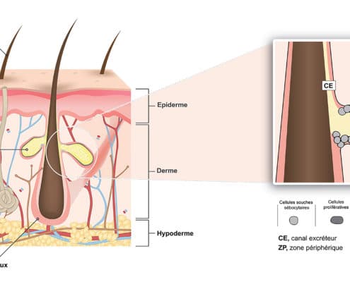
Physiology and functions of the sebaceous gland - Bioalternatives

Chapter 6 Worksheet Flashcards | Quizlet

21,931 Skin Anatomy Stock Photos, Pictures & Royalty-Free ...
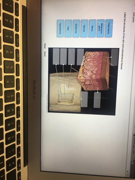
Solved Label the structure of the skin and subcutaneous ...
Structure Anatomy Human Hair Human Hair Stock Vector (Royalty ...
4.1 Layers of the Skin – Fundamentals of Anatomy and Physiology

Anatomy 2017- Unit 2 Label Parts of Skin Diagram Diagram ...
Solved Label the parts of the skin and subcutaneous tissue ...
Integumentary System - Laboratory Exercise (Anatomy and ...

BSC2085L - PARTS OF SKIN HW .docx - BSC2085L TISSUE HW LABEL ...
The subcutaneous layer: Anatomy, composition, and functions

Chapter 06 Lecture Outline - ppt download

label the parts of the skin using the word bank below ...
Post a Comment for "44 label the parts of the skin and subcutaneous tissue"