45 compound microscope diagram with labels
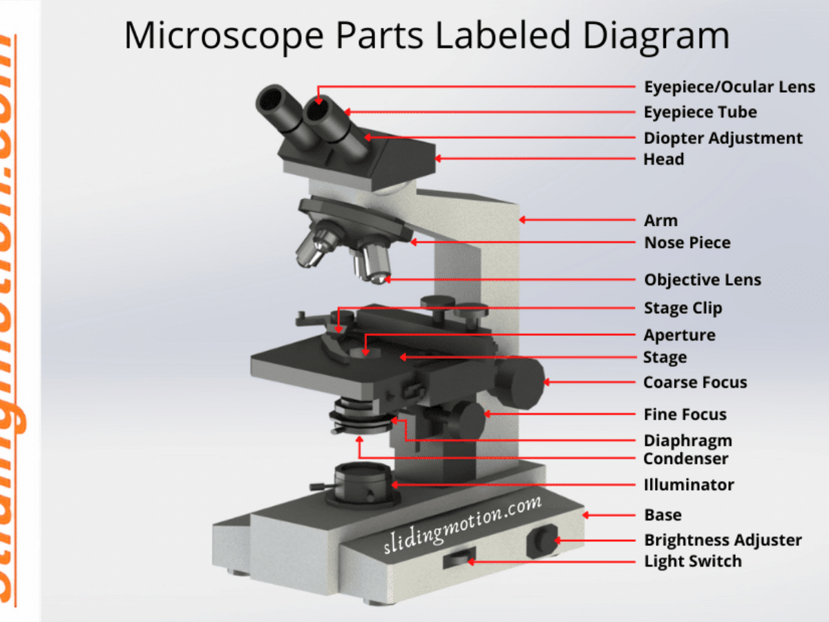
Compound Microscope Parts, Functions, and Labeled Diagram Nov 18, 2020 · The individual parts of a compound microscope can vary heavily depending on the configuration & applications that the scope is being used for. Common compound microscope parts include: Compound Microscope Definitions for Labels Eyepiece (ocular lens) with or without Pointer: The part that is looked through at the top of the compound microscope ... › Subaru_FA20E-FA20F_EnginesFA20E and FA20F Subaru Engines - australiancar.reviews The FA20E and FA20F engines have a cast aluminium alloy cylinder head with chain-driven double overhead camshafts per cylinder bank. The four valves per cylinder – two intake and two exhaust – were actuated by roller rocker arms which had built-in needle bearings that reduced the friction that occurred between the camshafts and the roller rocker arms.
16 Parts of a Compound Microscope: Diagrams and Video The 16 core parts of a compound microscope are: Head (Body) Arm Base Eyepiece Eyepiece tube Objective lenses Revolving Nosepiece (Turret) Rack stop Coarse adjustment knobs Fine adjustment knobs Stage Stage clips Aperture Illuminator Condenser Diaphragm Video: Parts of a compound Microscope with Diagram Explained

Compound microscope diagram with labels
Compound microscope - BiochemGems Figure: A labeled diagram of a compound microscope. Image formed by a compound microscope. The objective lens forms a true, inverted picture while the eyepiece functions as a basic magnifier that does not re-invert and generates a virtual image. The image always ends up inverted from the original. If we move the sample to the left, it will move ... Assignment Essays - Best Custom Writing Services Get 24⁄7 customer support help when you place a homework help service order with us. We will guide you on how to place your essay help, proofreading and editing your draft – fixing the grammar, spelling, or formatting of your paper easily and cheaply. assignmentessays.comAssignment Essays - Best Custom Writing Services Get 24⁄7 customer support help when you place a homework help service order with us. We will guide you on how to place your essay help, proofreading and editing your draft – fixing the grammar, spelling, or formatting of your paper easily and cheaply.
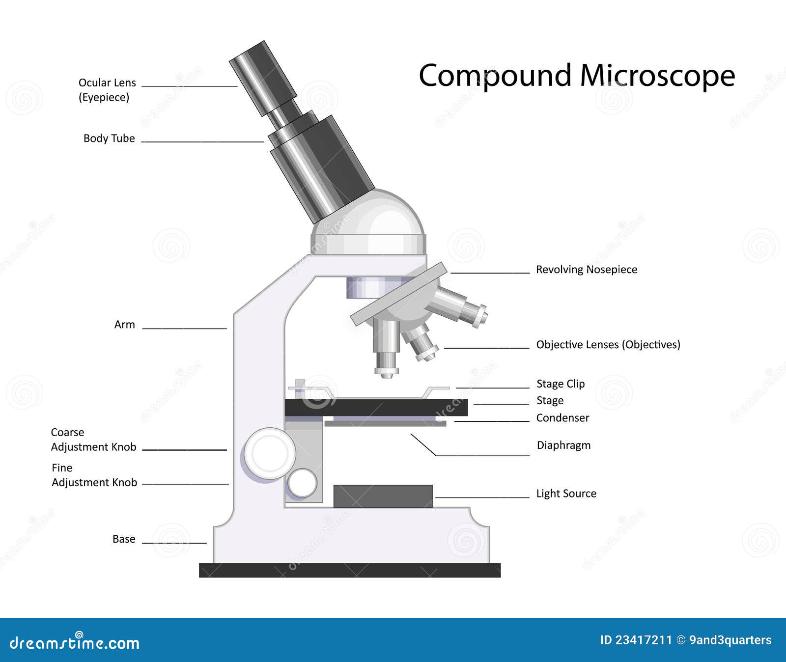
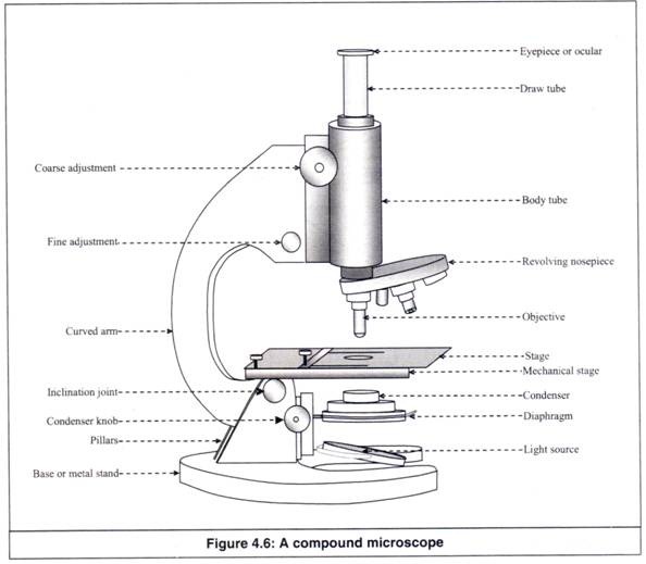
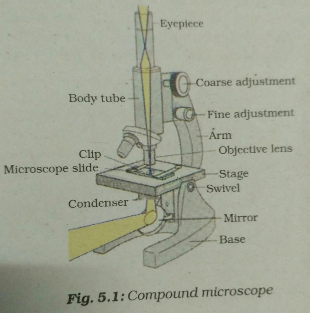
Compound microscope diagram with labels. Parts of a Compound Microscope - Labeled (with diagrams) Parts of a Compound Microscope - Labeled (with diagrams) A compound microscope is known as a high-power microscope that enables you to achieve a high level of magnification. Smaller specimens can be thoroughly viewed using a compound microscope. Let us take a look at the different parts of a compound microscope and understand each key component. How to draw compound of Microscope easily - step by step I will show you " How to draw compound of microscope easily - step by step "Please watch carefully and try this okay.Thanks for watching.....#microscopedrawi... Labelled Diagram of Compound Microscope The below mentioned article provides a labelled diagram of compound microscope. Part # 1. The Stand: The stand is made up of a heavy foot which carries a curved inclinable limb or arm bearing the body tube. The foot is generally horse shoe-shaped structure (Fig. 2) which rests on table top or any other surface on which the microscope in kept. Compound Microscope Parts - Labeled Diagram and their Functions What is a "compound microscope"? Labeled diagram of a compound microscope Major structural parts of a compound microscope Optical components of a compound microscope Eyepiece Eyepiece tube Objective lenses Nosepiece Specimen stage Coarse and fine focus knobs Rack stop Illuminator Condenser Abbe condenser Iris Diaphragm Condenser Focus Knob Summary
Parts of Stereo Microscope (Dissecting microscope) – labeled diagram ... If you would like to learn optical components of a compound microscope, please visit Compound Microscope Parts – Labeled Diagram and their Functions, and this article. How to use a stereo (dissecting) microscope. Follow these steps to put your stereo microscopes in work: 1. Set your microscope on a tabletop or other flat sturdy surface where ... High-entropy alloys | Nature Reviews Materials Jun 18, 2019 · High-entropy alloys have greatly expanded the compositional space for alloy design. In this Review, the authors discuss model high-entropy alloys with interesting properties, the physical ... Fluorescence - Wikipedia Fluorescence is the emission of light by a substance that has absorbed light or other electromagnetic radiation.It is a form of luminescence.In most cases, the emitted light has a longer wavelength, and therefore a lower photon energy, than the absorbed radiation.A perceptible example of fluorescence occurs when the absorbed radiation is in the ultraviolet … Microscope Types (with labeled diagrams) and Functions A compound microscope: Is used to view samples that are not visible to the naked eye Uses two types of lenses - Objective and ocular lenses Has a higher level of magnification - Typically up to 2000x Is used in hospitals and forensic labs by scientists, biologists and researchers to study micro organisms Compound microscope labeled diagram
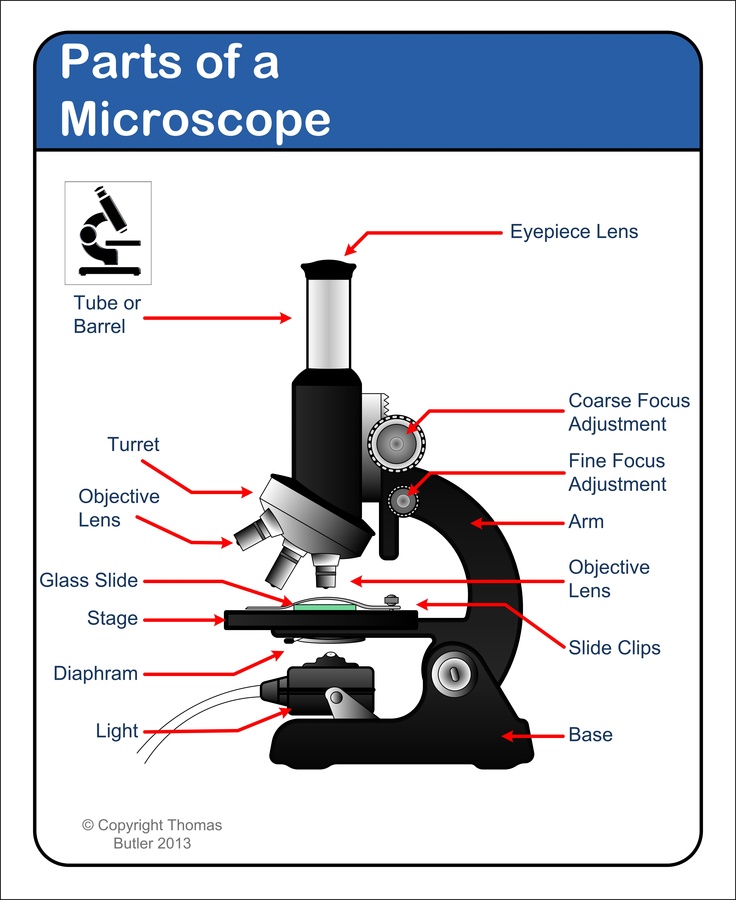
Microscope Parts and Functions Here are the important compound microscope parts... Eyepiece: The lens the viewer looks through to see the specimen. The eyepiece usually contains a 10X or 15X power lens. Diopter Adjustment: Useful as a means to change focus on one eyepiece so as to correct for any difference in vision between your two eyes. › createJoin LiveJournal Password requirements: 6 to 30 characters long; ASCII characters only (characters found on a standard US keyboard); must contain at least 4 different symbols; microscopeinternational.com › compound-microscopeCompound Microscope Parts, Functions, and Labeled Diagram Nov 18, 2020 · The individual parts of a compound microscope can vary heavily depending on the configuration & applications that the scope is being used for. Common compound microscope parts include: Compound Microscope Definitions for Labels Eyepiece (ocular lens) with or without Pointer: The part that is looked through at the top of the compound microscope ... FA20E and FA20F Subaru Engines - australiancar.reviews The FA20E and FA20F engines have a cast aluminium alloy cylinder head with chain-driven double overhead camshafts per cylinder bank. The four valves per cylinder – two intake and two exhaust – were actuated by roller rocker arms which had built-in needle bearings that reduced the friction that occurred between the camshafts and the roller rocker arms.
Microscope Parts and Functions
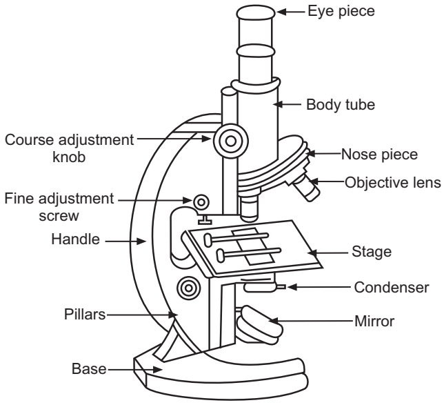
Diagram of a Compound Microscope - Biology Discussion Diagram of a Compound Microscope Article Shared by ADVERTISEMENTS: In this article we will discuss about:- 1. Essential Parts of Compound Microscope 2. Magnification of the Image of the Object by Compound Microscope 3. Resolution Power 4. Method for Studying Microbes 5. Measurement of the Size of Objects. Essential Parts of Compound Microscope:

Diagram of a Compound Microscope
Multiphoton Microscopy | Nikon’s MicroscopyU Two-photon excitation microscopy (also referred to as non-linear, multiphoton, or two-photon laser scanning microscopy) is an alternative to confocal and deconvolution microscopy that provides distinct advantages for three-dimensional imaging.In particular, two-photon excitation excels at imaging of living cells, especially within intact tissues such as brain slices, embryos, whole …

The Compound Microscope parts & how they work
Compound Microscope- Definition, Labeled Diagram, Principle, Parts, Uses Alternatively, the magnification of the compound microscope is given by: m = D/ fo * L/fe where, D = Least distance of distinct vision (25 cm) L = Length of the microscope tube fo = Focal length of the objective lens fe = Focal length of the eye-piece lens Parts of a Compound Microscope Eyepiece And Body Tube.

Compound Microscope- Definition, Labeled Diagram, Principle ...
Rolling back human pluripotent stem cells to an eight-cell ... - Nature Mar 21, 2022 · The development of a transgene-free, rapid and controllable method for producing eight-cell-like cells from human pluripotent stem cells provides a valuable resource to study early human ...

Can someone can send me diagram of this compound microscope ...
microscopewiki.com › simple-microscopeSimple Microscope - Diagram (Parts labelled), Principle ... Oct 01, 2022 · The basic difference between a simple microscope and compound microscope is the number of lenses used to magnify a specimen. While a simple microscope uses single lens to magnify an object, a compound microscope uses multiple lens to achieve higher order of magnification.

Draw a neat labelled diagram of a compound microscope class ...
Simple Microscope - Diagram (Parts labelled), Principle, Formula … Oct 01, 2022 · Simple Microscope Diagram with Labels Simple Microscope – Student Free Worksheet . Frequently Asked Questions . Q 1. How many lenses does a simple microscope have? ... The basic difference between a simple microscope and compound microscope is the number of lenses used to magnify a specimen. While a simple microscope uses single lens to ...

Compound Microscope Parts, Function, & Diagram | What is a ...
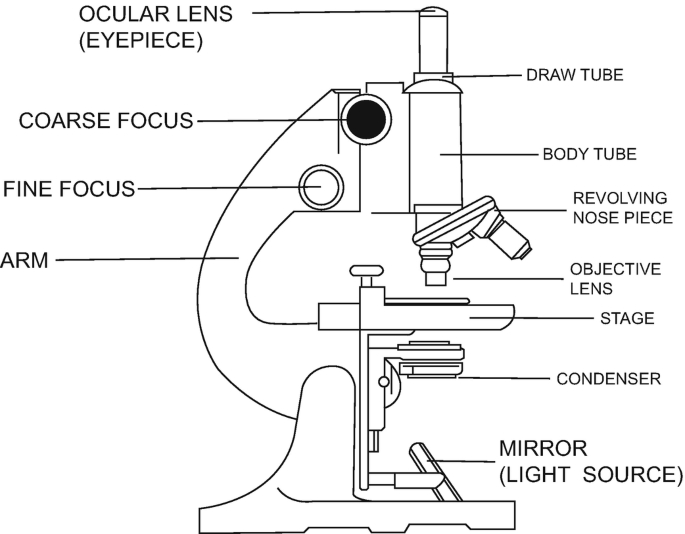
Compound Microscope - Diagram (Parts labelled), Principle and Uses Image : Labeled Diagram of compound microscope parts See: Labeled Diagram showing differences between compound and simple microscope parts Structural Components The three structural components include 1. Head This is the upper part of the microscope that houses the optical parts 2. Arm

Cell Drawing Microscope - Binocular Compound Microscope ...
Join LiveJournal Password requirements: 6 to 30 characters long; ASCII characters only (characters found on a standard US keyboard); must contain at least 4 different symbols;
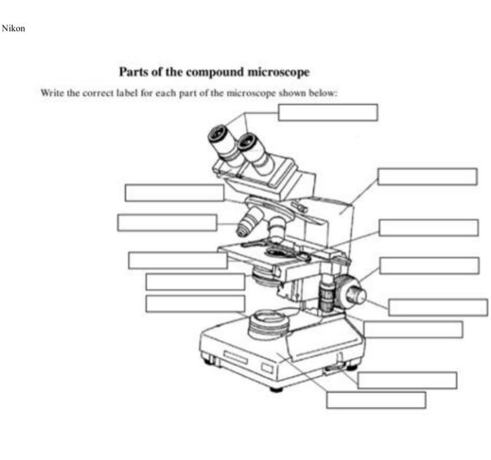
Solved Nikon Parts of the compound microscope Write the ...
rsscience.com › stereo-microscopeParts of Stereo Microscope (Dissecting microscope) – labeled ... Stereo microscopes (also called Dissecting microscope) are branched out from other light microscopes for the application of viewing "3D" objects. These include substantial specimens, such as insects, feathers, leaves, rocks, sand grains, gems, coins, and stamps, etc. Functionally, a stereo microscope is like a powerful magnifying glass.

The Microscope
Label a Compound Microscope Diagram | Quizlet Start studying Label a Compound Microscope. Learn vocabulary, terms, and more with flashcards, games, and other study tools. ... Label this. Illuminator Switch. Sets found in the same folder. AP II Ch. 24 Digestive Lab QUIZ. 10 terms. CWRN2016. Body planes Label. 9 terms. Hesi_Study.
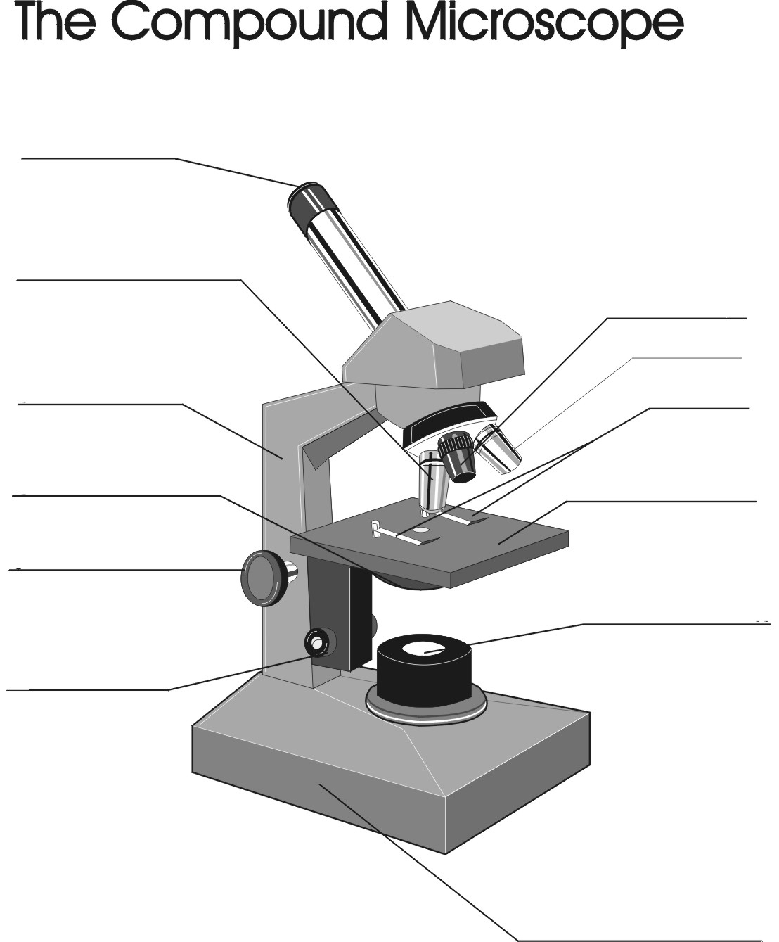
Remix of "The Compound Microscope"
Label the microscope — Science Learning Hub Use this interactive to identify and label the main parts of a microscope. Drag and drop the text labels onto the microscope diagram. eye piece lens diaphragm or iris coarse focus adjustment stage base fine focus adjustment light source high-power objective Download Exercise Tweet

Compound Microscope Labeled Diagram | Quizlet
A Study of the Microscope and its Functions With a Labeled Diagram ... A Study of the Microscope and its Functions With a Labeled Diagram To better understand the structure and function of a microscope, we need to take a look at the labeled microscope diagrams of the compound and electron microscope. These diagrams clearly explain the functioning of the microscopes along with their respective parts.

Draw a labelled diagram of a compound microscope.
› techniques › multi-photonMultiphoton Microscopy | Nikon’s MicroscopyU This is a valuable enhancement to the capability of the conventional microscope since ultraviolet wavelengths below approximately 300 nanometers are very problematic for regular microscope optics. Higher-order non-linear effects, such as four-photon absorption, have been experimentally demonstrated, although it is unlikely that these phenomena ...
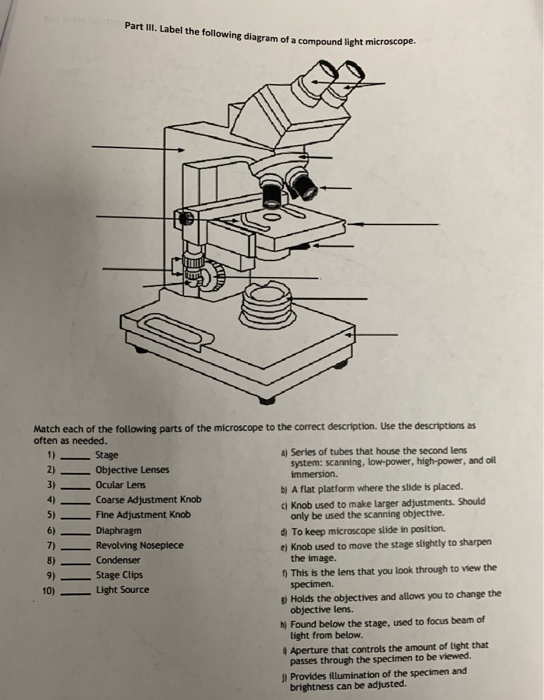
Solved Part III. Label the following diagram of a compound ...
Compound Microscope Labeled Diagram | Quizlet Compound Microscope Labeled Diagram | Quizlet Compound Microscope Labeled + − Flashcards Learn Test Match Created by meganplocher734 Terms in this set (14) Eyepiece/Ocular lens Contains the ocular lens Body tube A hollow cylinder that holds the eyepiece. Arm Part that supports the microscope. Stage Supports the slide or specimen

Drawn and lable the diagram of compound microscope and ...
Compound Microscope Diagram Labeled - microscope diagram tim s ... Compound Microscope Diagram Labeled - 17 images - light microscope diagram labeled micropedia, introduction to the light microscope flashcards easy, microscope diagram with name edusip, understanding the compound microscope parts and its,

Labeling the Parts of the Microscope | Microscope World Resources
Binocular Microscope Anatomy - Parts and Functions with a Labeled Diagram Now, I will discuss the details anatomy of the light compound microscope with the labeled diagram. Why it is called binocular: because it has two ocular lenses or an eyepiece on the head that attaches to the objective lens, this ocular lens magnifies the image produced by the objective lens. Binocular microscope parts and functions
Answered: Microscope Structure and Function… | bartleby
Compound Microscope: Definition, Diagram, Parts, Uses, Working ... - BYJUS The parts of a compound microscope can be classified into two: Non-optical parts Optical parts Non-optical parts Base The base is also known as the foot which is either U or horseshoe-shaped. It is a metallic structure that supports the entire microscope. Pillar The connection between the base and the arm are possible through the pillar. Arm
Compound Microscope Stock Illustrations – 736 Compound ...
assignmentessays.comAssignment Essays - Best Custom Writing Services Get 24⁄7 customer support help when you place a homework help service order with us. We will guide you on how to place your essay help, proofreading and editing your draft – fixing the grammar, spelling, or formatting of your paper easily and cheaply.
parts of microscope with diagram - Clip Art Library
Assignment Essays - Best Custom Writing Services Get 24⁄7 customer support help when you place a homework help service order with us. We will guide you on how to place your essay help, proofreading and editing your draft – fixing the grammar, spelling, or formatting of your paper easily and cheaply.

Difference between Simple and Compound Microscope ...
Compound microscope - BiochemGems Figure: A labeled diagram of a compound microscope. Image formed by a compound microscope. The objective lens forms a true, inverted picture while the eyepiece functions as a basic magnifier that does not re-invert and generates a virtual image. The image always ends up inverted from the original. If we move the sample to the left, it will move ...

How to draw compound of Microscope easily - step by step
Label Parts Of A Compound Microscope Teaching Resources | TPT

Living Environment Course

Simple Microscope Definition, Magnification, Parts And Uses
Cytology. Cytology. radiation used to illuminate the specimen ...
biology labeled microscope diagram - Clip Art Library

Simple Microscope - Diagram (Parts labelled), Principle ...
What is Compound Microscope? - Diagram, Function, Advantages

Compound Microscope Parts – Labeled Diagram and their ...
MICROBIO 16 Parts of a Compound Microscope with Diagram and ...

Parts of a Microscope with Their Functions – Microbe Online

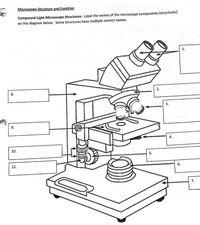
Activity 1: Name Me!Directions: Identify the parts of the ...

Compound Microscope Parts, Functions, and Labeled Diagram ...
The Compound Light Microscope Label the following parts on ...
Compound Microscope - Types, Parts, Diagram, Functions and ...
16 Basic Parts of Microscope, Function, Names & Labeled Diagram

Compound Microscope Parts – Labeled Diagram and their ...

Label the microscope — Science Learning Hub

Label Microscope Diagram - EnchantedLearning.com
Write any two parts of a compound microscope? | EduRev Class ...

easy compound microscope diagram - Clip Art Library

Compound Microscope Parts, Functions, and Labeled Diagram ...

2.1 " Compound Microscope" | Download Scientific Diagram

microscope | Types, Parts, History, Diagram, & Facts | Britannica
Exercise 1: Using a Compound Microscope | SpringerLink
Post a Comment for "45 compound microscope diagram with labels"